

Mycobacterium tuberculosis infection outside the lung usually results from hematogenous dissemination. Sometimes infection directly extends from an adjacent organ. Symptoms vary by organ or anatomic site but generally include fever, malaise, and weight loss. Diagnosis is most often based on cerebrospinal fluid or urine, or pleural, pericardial, or joint fluid testing (including molecular diagnostic tests) and culture. Evaluation for pulmonary TB is warranted if a pulmonary infection is suspected. Treatment is with combinations of antimicrobials for a duration of at least 6 months.
Tuberculosis (TB) properly refers only to disease caused by Mycobacterium tuberculosis (for which humans are the main reservoir). Although the lungs are most commonly the initial site of infection, the disease can spread to many organs. (For details on the organism, pathophysiology, and pulmonary disease, see Tuberculosis.)
Pleural TB (TB empyema)
Empyema in extrapulmonary TB results from chronic, purulent infection of the pleural space in patients with a high volume of tubercle bacilli. Empyema often occurs because of direct rupture of a contiguous pulmonary cavity into the pleura.
The clinical presentation is usually grossly purulent pleural fluid and marked pleural thickening on imaging, and because of the severity of both, surgical drainage and prolonged antituberculous chemotherapy may be needed.
Pericardial TB (TB pericarditis)
Pericardial infection may develop from foci in mediastinal lymph nodes or from pleural TB. In some high-prevalence parts of the world, TB pericarditis is a common cause of heart failure.
Patients may have a pericardial friction rub, pleuritic and positional chest pain, or fever. Pericardial tamponade may occur, causing dyspnea, neck vein distention, paradoxical pulse, muffled heart sounds, and possibly hypotension.
Tuberculous lymphadenitis (scrofula)
Tuberculous lymphadenitis typically involves the lymph nodes in the posterior cervical and supraclavicular chains. Infection in these areas is thought to be due to contiguous spread from intrathoracic lymphatics or from infection in the tonsils and adenoids. Mediastinal and hilar lymph nodes are also commonly enlarged as a part of primary pulmonary disease, especially in children (3). The combination of the initial lung parenchymal area of infection (Ghon focus) and affected lymph nodes is called a Ghon complex, and a calcified Ghon complex is known as a Ranke complex.
Cervical tuberculous lymphadenitis is characterized by progressive swelling of the affected nodes. In advanced cases, nodes may become inflamed and tender; the overlying skin may break down, resulting in a draining fistula.
Scrofula was called "King's evil" during the 13th through 18th centuries in England and France, and it was widely believed that the lesions could be healed by the touch of a member of royalty (4).
Miliary TB
Miliary TB is a severe, potentially fatal form of M. tuberculosis infection (1). Also known as generalized hematogenous TB, miliary TB develops when a tuberculous lesion erodes into a blood vessel, disseminating millions of tubercle bacilli into the bloodstream and throughout the body. Miliary TB is characterized by the presence of tuberculous nodules (tuberculomas) in multiple organs. The so-called millet seed–like appearance of these nodules on gross pathology or imaging is what is termed "miliary." The lungs and bone marrow are most often affected, but any site may be involved. Uncontrolled massive dissemination can occur during primary infection or after reactivation of a latent focus.
Miliary TB is most common among:
Children < 4 years old
Immunocompromised people (especially those with HIV infection)
Older adults
Symptoms of miliary TB include fever, chills, weakness, malaise, weight loss, and often progressive dyspnea. Intermittent dissemination of tubercle bacilli may lead to a prolonged fever of unknown origin (FUO).
Choroidal tubercles (small, yellowish inflammatory lesions in the choroid layer of the eye) are pathognomonic if present.
Bone marrow involvement may cause anemia, thrombocytopenia, or a leukemoid reaction.
In untreated patients with miliary TB and immunosuppression, disease often progresses rapidly to acute respiratory failure or acute respiratory distress syndrome (ARDS) and multi-organ dysfunction; mortality can consequently be high.
Meningeal TB (TB meningitis)
TB meningitis often occurs in the absence of infection at other extrapulmonary sites. In the United States, TB meningitis is most common among older adults and immunocompromised patients. In areas of the world where TB is common among children, TB meningitis usually occurs in newborns and children through age 5.
Meningitis is the most serious manifestation of TB and is associated with high morbidity as well as mortality in approximately 29% of cases (2). TB meningitis is one of the forms of severe TB that can be prevented in children by BCG vaccine.
Symptoms include low-grade fever, unremitting headache, nausea, and drowsiness, which may progress to stupor and coma. Kernig and Brudzinski signs may be positive. Focal neurologic symptoms suggest a tuberculoma.
Because the early signs are non-specific, it is important to consider the diagnosis early in any patient with known TB exposure, infection, or disease, including a prior history of TB, and in all people with compatible symptoms who are from high TB-prevalence locations.
The clinical course of TB meningitis progresses through the following stages:
Stage 1: Clear sensorium with abnormal cerebrospinal fluid (CSF) examination
Stage 2: Drowsiness or stupor with focal neurologic signs
Stage 3: Coma
Potential complications include stroke, which may result from thrombosis of a major cerebral vessel, and coma.
Gastrointestinal TB
Because the entire gastrointestinal (GI) mucosa resists TB invasion, infection requires prolonged exposure and enormous inocula. GI TB is very unusual in countries where bovine TB is rare (eg, because of milk pasteurization and routine TB testing of cattle).
Ulcers of the mouth and oropharynx may develop from eating M. bovis–contaminated dairy products; primary lesions may also occur in the small bowel. Intestinal invasion generally causes hyperplasia and an inflammatory bowel syndrome with pain, diarrhea, obstruction, and hematochezia. It may also mimic appendicitis. Ulceration and fistulas are possible.
Hepatic TB
Hepatic TB is common among patients with advanced pulmonary TB and widely disseminated or miliary TB. However, the liver generally heals without sequelae when the principal infection is treated.
TB in the liver occasionally spreads to the gallbladder, leading to obstructive jaundice.
Genitourinary TB
TB infection of the kidneys may manifest as pyelonephritis (eg, fever, back or flank pain, pyuria) without the usual urinary pathogens on routine culture (sterile pyuria).
Infection commonly spreads to the bladder and, in men, to the prostate, seminal vesicles, or epididymis, causing an enlarging scrotal mass. Infection may spread to the perinephric space and down the psoas muscle, sometimes causing an abscess on the anterior thigh.
In women, salpingo-oophoritis can occur after menarche, when the fallopian tubes become vascular. Symptoms include chronic pelvic pain and sterility or ectopic pregnancy due to tubal scarring.
Peritoneal TB (TB peritonitis)
Peritoneal infection represents seeding from abdominal lymph nodes or from salpingo-oophoritis. TB peritonitis is particularly common among people with alcohol use disorder who have cirrhosis.
Symptoms may be mild, with fatigue, abdominal pain, and tenderness, or severe enough to mimic those of acute abdomen.
Cutaneous TB
Cutaneous TB can take many characteristic forms, including scrofuloderma, lupus vulgaris, and verrucosa cutis (5). Symptoms range from papules, nodules, and wart-like lesions to chronic ulcers, sinus tracts, and abscesses.
Scrofuloderma results from direct extension of an underlying TB focus (eg, a regional lymph node, an infected bone or joint) to the overlying skin, forming ulcers and sinus tracts.


Patients with scrofuloderma have painless, firm, subcutaneous nodules (top arrow) that eventually enlarge and form ulcers and sinus tracts (bottom arrow).
Lupus vulgaris results from hematogenous or lymphogenous dissemination to the skin from an extracutaneous focus in a sensitized patient.

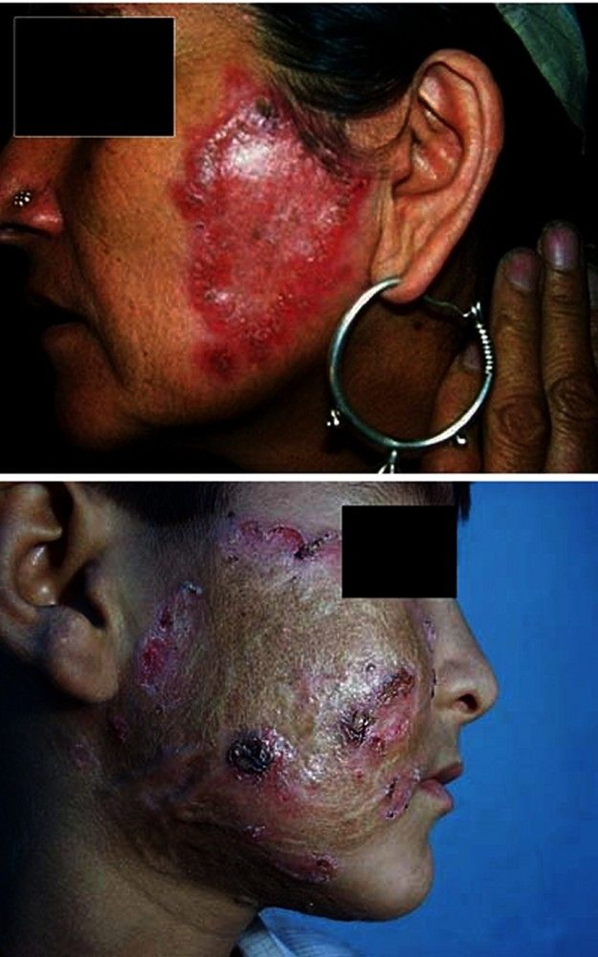
The top image shows irregularly shaped red plaques in a patient with lupus vulgaris, which is a form of cutaneous tuberculosis.
The bottom image shows an erythematous plaque with central clearing and an outermost raised erythematous border in a patient with lupus vulgaris.
© Springer Science+Business Media
Tuberculosis verrucosa cutis (prosector's wart) occurs after exogenous direct inoculation of the mycobacteria into the skin of a previously sensitized patient who has moderate to high immunity against the bacilli. Rashes are typically erythematous and papulonodular in appearance.
Rarely, TB develops on abraded skin in patients with cavitary pulmonary TB.


This photo shows the rash of tuberculosis verrucosa cutis (prosector's wart) on the dorsal surface of the hand. The rash consists of small, erythematous, papular nodules.
DoubleVision/SCIENCE PHOTO LIBRARY
Musculoskeletal TB
Weight-bearing joints are most commonly involved, but bones of the wrist, hand, and elbow may also be affected, especially after injury.
Pott disease is spinal infection, which begins in a vertebral body and often spreads to adjacent vertebrae, with narrowing of the disc space between them. Untreated, the vertebrae may collapse, possibly impinging on the spinal cord. Symptoms include progressive or constant pain in involved bones and chronic or subacute arthritis (usually monoarticular). In Pott disease, spinal cord compression causes neurologic deficits, including paraplegia; paravertebral swelling may result from an abscess.
Tubercle bacilli may spread to tendon sheaths (tuberculous tenosynovitis) by direct extension from adjacent lesions in bone or hematogenously from any infected organ.
Other sites of TB infection
TB may infect the wall of a blood vessel and can cause the aorta to rupture.
Adrenal involvement, leading to Addison disease, was common but now is rare.
General references
1. Sharma SK, Mohan A, Sharma A, Mitra DK. Miliary tuberculosis: new insights into an old disease. Lancet Infect Dis. 2005;5(7):415-430. doi:10.1016/S1473-3099(05)70163-8
2. Seid G, Alemu A, Dagne B, Gamtesa DF. Microbiological diagnosis and mortality of tuberculosis meningitis: Systematic review and meta-analysis. PLoS One. 2023;18(2):e0279203. Published 2023 Feb 16. doi:10.1371/journal.pone.0279203
3. Mahomed N, Kilborn T, Smit EJ, et al. Tuberculosis revisted: classic imaging findings in childhood. Pediatr Radiol. 2023;53(9):1799-1828. doi:10.1007/s00247-023-05648-z
4. Grzybowski S, Allen EA. History and importance of scrofula. Lancet. 1995;346(8988):1472-1474. doi:10.1016/s0140-6736(95)92478-7
5. Nguyen KH, Alcantara CA, Glassman I, et al. Cutaneous Manifestations of Mycobacterium tuberculosis: A Literature Review. Pathogens. 2023;12(7):920. Published 2023 Jul 8. doi:10.3390/pathogens12070920
Diagnosis of Extrapulmonary TB
Acid-fast bacillus (AFB) microscopy; molecular tests (nucleic acid amplification testing [NAAT]); and mycobacterial culture of fluid and tissue samples
Imaging studies of affected organs
Tuberculin skin test (TST) or interferon-gamma release assay (IGRA)
The diagnosis of extrapulmonary TB is generally similar to that for pulmonary TB (see Diagnosis of TB) and includes chest imaging, TST or IGRA, molecular tests, AFB microscopy, and mycobacterial culture (1).
NAAT and microscopic examination of specimens with AFB staining are also recommended but based on lower-quality evidence. Mycobacterial cultures of affected body fluids (cerebrospinal fluid, urine, or pleural, pericardial, or joint fluid) and tissue biopsies are typically performed. Blood culture results are positive in approximately 50% of patients with disseminated (miliary) TB; such patients are often immunocompromised, often because of HIV infection.


This image is a light micrograph of an acid-fast–stained sputum sample containing M. tuberculosis bacteria. The red color that remains after acid alcohol treatment indicates they are acid-fast.
CDC/SCIENCE PHOTO LIBRARY
However, cultures and smears of body fluids and tissues are often negative because few organisms are present; in such cases, NAAT, when available, may be particularly helpful. NAAT provides rapid results. This testing can be performed on fresh fluid or biopsy samples and on fixed tissue (eg, if TB was not suspected during a surgical procedure and testing of fresh tissue was not done) (1). A positive NAAT result almost always supports a TB diagnosis because NAAT has a high specificity for TB detection (typically > 95%); however, a negative result does not exclude TB in most cases because NAAT has low sensitivity (often < 90% but may depend on specimen tested) (1).
Other findings in body fluids depend on microscopic or other testing. Typically, lymphocytosis is present in body fluids. A very suggestive finding in the cerebrospinal fluid is a glucose level < 50% of that in serum and an elevated protein level.
If all tests are negative and miliary TB is still a concern, biopsies of the bone marrow and the liver are performed. If TB is highly suspected based on other features (eg, granuloma seen on biopsy, positive TST or IGRA result plus unexplained lymphocytosis in pleural fluid or cerebrospinal fluid), treatment should usually proceed despite inability to demonstrate TB organisms.
Chest radiography and other imaging (eg, high-resolution CT) can also provide helpful diagnostic information (2). Chest radiograph may show signs of primary or active TB; in miliary TB, it shows thousands of 2- to 3-mm interstitial nodules evenly distributed through both lungs.

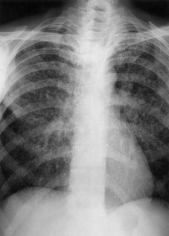
This chest radiograph show findings suggesting miliary tuberculosis (TB), including almost innumerable, tiny (eg, 2- to 3-mm), interstitial nodules distributed through both lungs.
Other imaging tests are done based on clinical findings. Abdominal or genitourinary involvement usually requires CT or ultrasonography; renal lesions are often visible. Bone and joint involvement requires CT or MRI; MRI is preferable for spinal disease because of the potential for soft-tissue involvement.
Diagnosis references
1. Lewinsohn DM, Leonard MK, LoBue PA, et al. Official American Thoracic Society/Infectious Diseases Society of America/Centers for Disease Control and Prevention Clinical Practice Guidelines: Diagnosis of Tuberculosis in Adults and Children. Clin Infect Dis. 2017;64(2):111-115. doi:10.1093/cid/ciw778
2. Rodriguez-Takeuchi SY, Renjifo ME, Medina FJ. Extrapulmonary Tuberculosis: Pathophysiology and Imaging Findings. Radiographics. 2019;39(7):2023-2037. doi:10.1148/rg.2019190109
Treatment of Extrapulmonary TB
Antibiotics
Sometimes glucocorticoids
Sometimes surgery
Treatment with antibiotics follows standard regimens and principles of TB management (see First-line medications for TB). Six to 9 months of therapy are usually adequate for most extrapulmonary sites except the meninges, which require treatment for 9 to 12 months.
Drug resistance is a major concern; it is increased by poor adherence, use of monotherapy, and inadequate susceptibility testing.
Glucocorticoids are often used to manage TB meningitis.
The United States Centers for Disease Control and Prevention (CDC) does not recommend the routine use of glucocorticoids for TB pericarditis that is not constrictive (1). However, glucocorticoids may prevent constriction in patients who are at risk.
Glucocorticoids may be used for meningitis and TB pericarditis (even when not constrictive) in patients with immune reconstitution inflammatory syndrome (IRIS).
Surgery is required for the following:
Drainage of fluid collections (eg, empyema, cardiac tamponade, central nervous system abscess)
Repair of bronchopleural fistulas
Resection of infected bowel
Decompression of masses encroaching on the spinal cord
Surgical debridement is sometimes needed in Pott disease to correct spinal deformities or to relieve cord compression if there are neurologic deficits or pain persists; fixation of the vertebral column by bone graft is required in only the most advanced cases.
Surgery is usually not necessary for TB lymphadenitis except for diagnostic purposes.
Treatment reference
1. Nahid P, Dorman SE, Alipanah N, et al. Official American Thoracic Society/Centers for Disease Control and Prevention/Infectious Diseases Society of America Clinical Practice Guidelines: Treatment of Drug-Susceptible Tuberculosis. Clin Infect Dis. 2016;63(7):e147-e195. doi:10.1093/cid/ciw376
Key Points
Tuberculosis can spread from the lungs through the bloodstream to many sites.
Symptoms depend on the affected organ but typically include fever, malaise, and weight loss.
TST and IGRA may suggest the presence of extrapulmonary TB but are not confirmatory.
Diagnosis based on identification of bacilli in infected fluid or tissue by microscopic examination and culture and/or molecular methods (nucleic acid amplification tests).
Treat with multiple medications for several months and sometimes with surgery.
Drug resistance is a major concern.


