


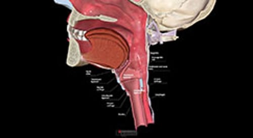
The larynx contains the vocal folds (also referred to as vocal cords) and serves as the opening to the tracheobronchial tree. Laryngeal disorders include, but are not limited to the following:
Other disorders that affect the larynx include acute laryngotracheobronchitis (croup), epiglottitis, and laryngomalacia (see table ). For removal of a foreign body via the Heimlich maneuver, see Clearing and Opening the Upper Airway.
Many laryngeal disorders cause dysphonia, which is impairment of the voice. Dysphonia can include a change in the sound of the voice (eg, hoarseness), its stability, or the effort required to produce it, among other features. A persistent change in the voice lasting ≥ 2 weeks requires visual examination of the vocal folds, including their mobility (1). Although the voice changes with advancing age, becoming breathy and aperiodic (ie, irregular vibration causing a rough or creaky sound), acute or prominent changes in older patients should not be presumed to result from aging, and evaluation is required (1).
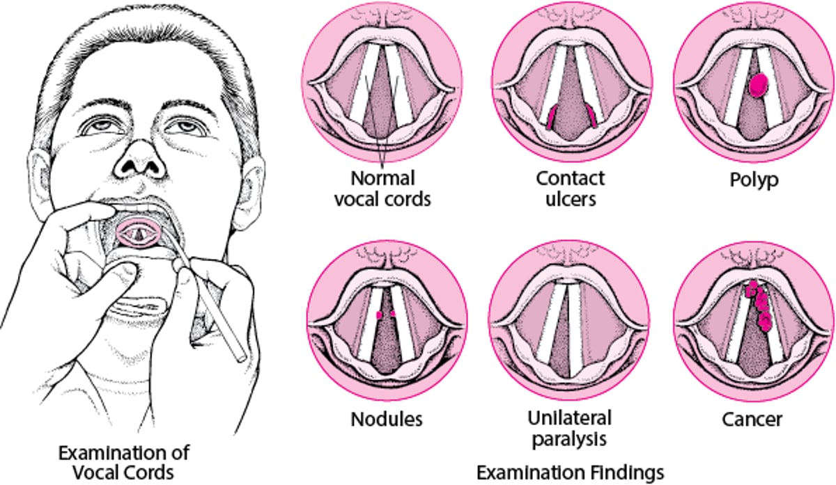
The examiner should take precautions to prevent respiratory infectious diseases as appropriate. The voice should be assessed and recorded, particularly if surgical procedures are planned. Examination of the larynx includes external inspection and palpation of the neck and internal visualization of the epiglottis, false vocal folds, true vocal folds, arytenoids, pyriform sinuses, and subglottic region. Examination can be accomplished by indirect mirror examination (see figure ), flexible laryngoscopy, or rigid transoral laryngoscopy in the outpatient setting with topical anesthetics, as needed. Rigid operative laryngoscopy under general anesthesia provides the most thorough anatomic examination of the vocal folds, allowing
Visualization of the under surfaces
Assessment of passive mobility when immobilized by either paralysis or fixation
Biopsy
Laryngeal Disorders
When relaxed, the vocal folds normally form a V-shaped opening that allows air to pass freely through to the trachea. The folds open during inspiration and close during swallowing or speech. When a mirror is held in the back of a patient’s mouth, the vocal folds can often be seen and checked for disorders, such as contact ulcers, polyps, nodules, paralysis, and cancer. Paralysis may affect one (unilateral) or both vocal folds (bilateral—not shown). |

Reference
1. Stachler RJ, Francis DO, Schwartz SR, et al. Clinical Practice Guideline: Hoarseness (Dysphonia) (Update) [published correction appears in Otolaryngol Head Neck Surg. 2018 Aug;159(2):403. doi: 10.1177/0194599818766900.]. Otolaryngol Head Neck Surg. 2018;158(1_suppl):S1-S42. doi:10.1177/0194599817751030


