


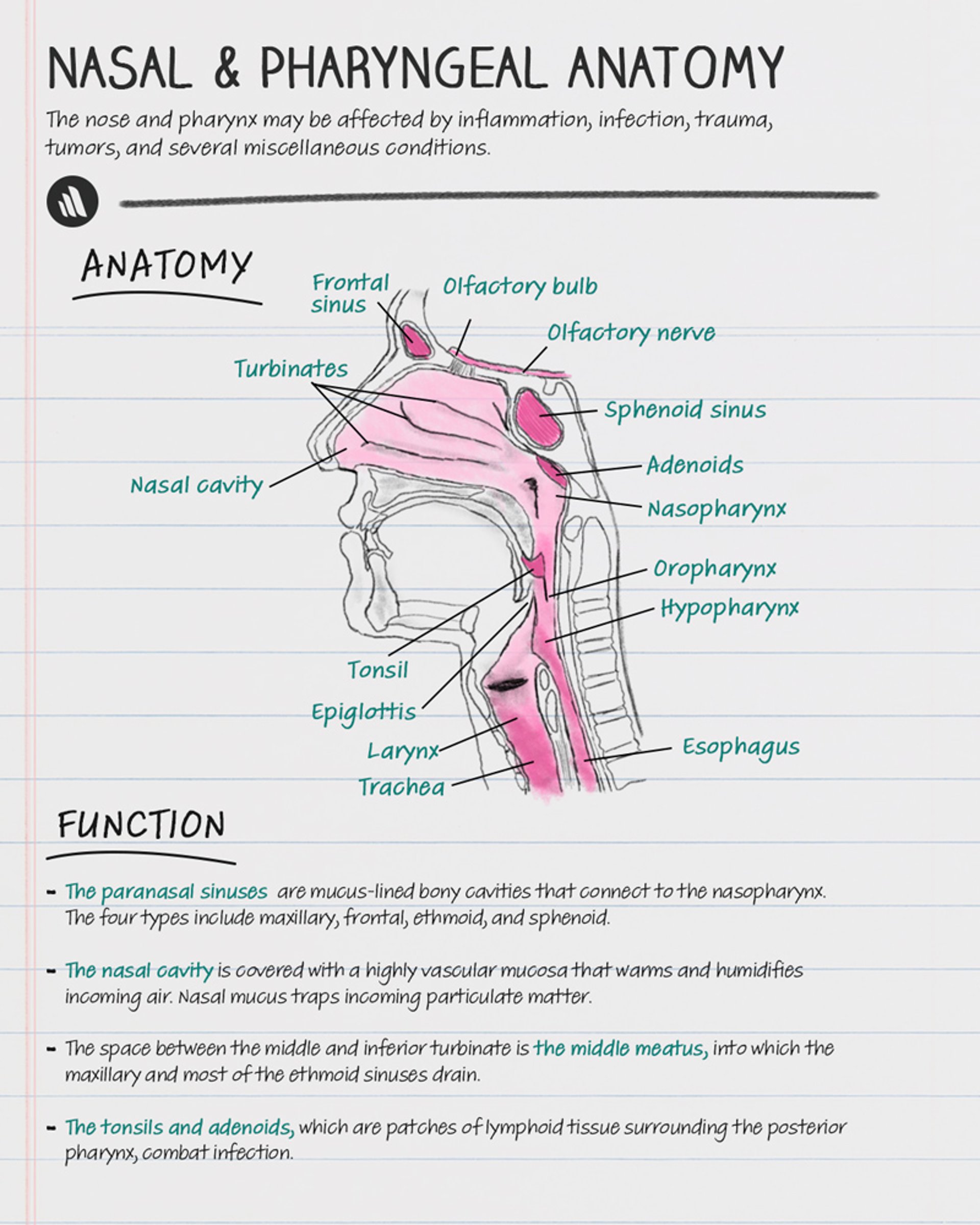
The nose and pharynx (consisting of the nasopharynx, oropharynx, and hypopharynx) may be affected by inflammation, infection, trauma, tumors, and several miscellaneous conditions.
Nasal and Pharyngeal Anatomy
Throat
The uvula hangs in the midline at the far end of the soft palate. It varies greatly in length. A long uvula and loose or excess velopharyngeal tissue may cause snoring and occasionally contribute to obstructive sleep apnea.
Tonsils and adenoids are patches of lymphoid tissue surrounding the posterior pharynx in an area termed Waldeyer’s ring. Their role is to combat infection.
The larynx is discussed in Laryngeal Disorders.

Copyright © 2023 Merck & Co., Inc., Rahway, NJ, USA and its affiliates. All rights reserved.
Nose
The nasal cavity is covered with a highly vascular mucosa that warms and humidifies incoming air. Each lateral wall of the cavity has three turbinates, which are bony shelves that increase the surface area, thus providing more effective heat and moisture exchange. Nasal mucus traps incoming particulate matter. The space between the middle and inferior turbinate is the middle meatus, into which the maxillary and most of the ethmoid sinuses drain. Polyps may develop between the turbinates, often in association with chronic sinusitis, asthma, allergy, aspirin use, and cystic fibrosis.
Sinuses
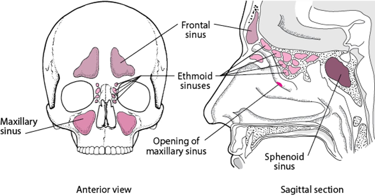
The paranasal sinuses are mucus-lined bony cavities that connect to the nasopharynx. There are 4 types, named based on location within the skull:
Maxillary
Frontal
Ethmoid
Sphenoid
They are located in the facial and cranial bones (see figure ). The physiologic role of the paranasal sinuses may include reducing skull weight and improving vocal resonance. They may also have immunologic functions involving the production of nitric oxide, which enhances local defense against pathogens and improves oxygen uptake in the respiratory tract (1).
Paranasal sinuses

Anatomy reference
1. Keir J. Why do we have paranasal sinuses?. J Laryngol Otol. 2009;123(1):4-8. doi:10.1017/S0022215108003976
Evaluation of the Nose and Pharynx
Examination of the nose and pharynx is part of every general otorhinolaryngologic examination.
History
General information includes use of alcohol or tobacco (both major risk factors for head and neck cancer) and systemic symptoms, such as fever and weight loss.
Oropharyngeal symptoms include
Mouth and throat pain
Mouth and throat ulcers
Difficulty swallowing or speaking
Nasal and sinus symptoms include
Congestion (noting presence and duration)
Sneezing
Nasal discharge (anterior or posterior/postnasal drip)
Loss of smell and/or taste
Bleeding from the nose
Physical examination
Some otorhinolaryngologic clinicians use a head-mounted light. However, because the light cannot be precisely aligned on the axis of vision, it is difficult to avoid shadowing in narrow areas (eg, nasal cavity). Illumination is better if the head-mounted mirror is convex; the clinician looks through a hole in the center of the mirror, so the illumination is always on-axis. The head mirror reflects light from a source placed behind the patient and slightly to one side and requires practice to use effectively.
The nose is examined using a nasal speculum, which is inserted into the nasal cavity with the two blades open in an anteroposterior (or slightly oblique) direction, not pressing against the septum. The clinician notes any crusting, discharge, septal deviation, or perforation; whether mucosa, especially the turbinates, are erythematous, boggy, or swollen; and presence of polyps. The skin over the frontal and maxillary sinuses is examined for erythema and tenderness, suggesting sinus inflammation.
If necessary, the nasopharynx and hypopharynx can be examined with a flexible nasopharyngoscope. A topical anesthetic (eg, lidocaine 4%) is sprayed in the nose and throat, and the nose is also sprayed with a decongestant (eg, phenylephrine 0.5%). After several minutes, the scope is gently passed through the nares, and the nasal cavity, hypopharynx, and larynx are inspected. A nasal endoscopic examination can also be done using a rigid scope, which provides higher-resolution views inside the nose but requires skill to use without causing the patient discomfort.
Alternatively, a mirror may be used to visualize the pharynx. A topical pharyngeal anesthetic for the throat is required for this examination. The mirror should be warmed before use to avoid fogging. A small mirror is used for the nasopharynx. It is held just below the uvula, angling upward; the tongue is pushed down with a tongue blade. A larger mirror is used for the hypopharynx and larynx. The tongue is retracted by grasping it with a gauze pad, and the mirror is placed against the soft palate, angling downward.
Neck examination consists of inspection and palpation for masses. If masses are found, the clinician notes whether they are tender, fluctuant, firm, or stony hard and whether they are mobile or fixed. Masses caused by infection are tender and mobile, whereas cancers tend to be nontender, hard, and fixed. Particular attention is paid to the cervical lymph nodes and thyroid and parotid glands.
Drug Information for the Topic


