

Actinic keratoses are precancerous changes in skin cells (keratinocytes) that are a frequent consequence of many years of sun exposure. Diagnosis is clinical. Treatment typically includes lesion-directed or field-directed therapy.
The prevalence of actinic keratoses is high and increases with age. They are considered intraepidermal keratinocyte neoplasms (1). Actinic keratoses are considered premalignant lesions that have a significant risk of progression to squamous cell carcinoma. The estimated rate of progression for an individual actinic keratosis to a squamous cell carcinoma varies widely, but consensus estimates most often range from < 1% to 10% (2, 3). Actinic keratoses that do not progress to squamous cell carcinoma may regress or persist as actinic keratoses. Lesions that regress may subsequently recur.
In addition to many years of sun exposure, other risk factors for actinic keratoses include older age, underlying immunosuppression, having blond or red hair, blue eyes, and light skin.
General references
1. Ibrahim SF, Brown MD. Actinic keratoses: a comprehensive update. J Clin Aesthet Dermatol. 2009;2(7):43-48
2. Dodds A, Chia A, Shumack S. Actinic keratosis: rationale and management. Dermatol Ther (Heidelb). 2014;4(1):11-31. doi:10.1007/s13555-014-0049-y
3. Werner RN, Sammain A, Erdmann R, et al. The natural history of actinic keratosis: a systematic review. Br J Dermatol. 2013;169(3):502-518. doi:10.1111/bjd.12420
Symptoms and Signs of Actinic Keratoses
Actinic keratoses often have adherent scales and are sometimes more easily felt than seen. Actinic keratoses can appear thickened or hypertrophic and sometimes form a cutaneous horn. They may be pink, red, or, less commonly, gray or brown. Lesions frequently develop in sun-exposed areas (eg, balding scalp, face, lateral neck, distal upper or lower extremities). Diffuse involvement of the lip is called actinic cheilitis.
Diagnosis of Actinic Keratoses
History and physical examination
Rarely, biopsy (for suspicious lesions)
The diagnosis of actinic keratoses is often based on visual and tactile examination; lesions feel rough and scaly on palpation. They should be differentiated from seborrheic keratoses, which increase in number and size with age. Seborrheic keratoses tend to appear waxy and stuck-on but can take on an appearance similar to that of actinic keratoses. Close inspection usually reveals distinguishing characteristics of the lesion. An actinic keratosis can also be distinguished from a seborrheic keratosis by the rough, gritty feel of the scale and the erythema. Unlike actinic keratoses, seborrheic keratoses also occur on non–sun-exposed areas of the body and are not precancerous.
Lesions that are atypical (ie, indurated or thickened, size > 1 cm, rapidly enlarging, bleeding, ulcerated) or fail to respond to standard therapy should be biopsied to exclude squamous cell carcinoma (1).

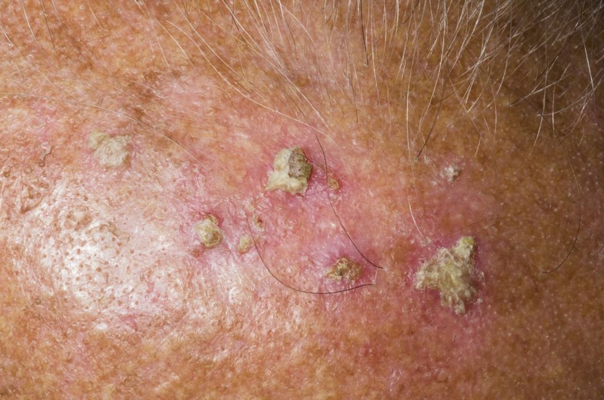
This photo shows an irregular erythematous patch with adherent yellowish tan scale.
DR P. MARAZZI/SCIENCE PHOTO LIBRARY
Diagnosis reference
1. Du Thanh A, Guillot B. Kératoses actiniques : quand biopsier ? [Actinic keratosis: when is a skin biopsy necessary?]. Eur J Dermatol. 2012 Dec;22 Suppl 1:13-6. French. doi: 10.1684/ejd.2012.1874
Treatment of Actinic Keratoses
Lesion-directed or field-directed therapy
Treatment options depend on the number of lesions, their location, extent of photodamage, and patient preference, but they are typically divided into either lesion- or field-directed therapy.
In lesion-directed therapy, individual lesions are physically removed. This approach is preferable for patients with only a few actinic keratoses. Cryosurgery (freezing with liquid nitrogen) is the most common lesion-directed therapy. Curettage (scraping with a curette) is an alternative. Lesion-directed therapies have the benefit of being one-time, in-office procedures, but they may not address subclinical changes and have a higher risk of scarring.
In field-directed therapy, topical treatments are applied to larger, more diffuse areas of involvement (1). This approach is appropriate for patients with multiple actinic keratoses and evidence of chronic actinic damage in adjacent areas (ie, field cancerization). First-line treatments include topical fluorouracil (5-FU) cream and imiquimod cream. Alternatives include diclofenac gel and tirbanibulin ointment.). This approach is appropriate for patients with multiple actinic keratoses and evidence of chronic actinic damage in adjacent areas (ie, field cancerization). First-line treatments include topical fluorouracil (5-FU) cream and imiquimod cream. Alternatives include diclofenac gel and tirbanibulin ointment.
5-FU inhibits thymidylate synthetase, limiting DNA synthesis and causing death of damaged cells. 5-FU 5% cream is applied 2 times a day for 3 to 4 weeks. One randomized trial showed 5-FU to be the most effective treatment for actinic keratoses compared with three other field-directed treatments (2). Another randomized trial suggested that combining 5% FU cream with 0.005% calcipotriol may enhance efficacy of 5-FU (3).
Imiquimod is an immune response modifier that stimulates local cytokine induction, resulting in a brisk local inflammatory reaction. A randomized trial showed that Imiquimod 5% cream applied 2 times a week for 16 consecutive weeks is effective and safe (Imiquimod is an immune response modifier that stimulates local cytokine induction, resulting in a brisk local inflammatory reaction. A randomized trial showed that Imiquimod 5% cream applied 2 times a week for 16 consecutive weeks is effective and safe (4).
Diclofenac is a nonsteroidal anti-inflammatory medication that inhibits both cyclooxygenase and upregulation of the arachidonic acid cascade. Diclofenac 3% (in a 2.5% hyaluronan gel) is applied 2 times a day for 60 to 90 days. Its use is limited by its low efficacy (Diclofenac is a nonsteroidal anti-inflammatory medication that inhibits both cyclooxygenase and upregulation of the arachidonic acid cascade. Diclofenac 3% (in a 2.5% hyaluronan gel) is applied 2 times a day for 60 to 90 days. Its use is limited by its low efficacy (5).
Tirbanibulin is a microtubule inhibitor that inhibits tubulin polymerization and Src kinase signaling, inducing death of damaged cells. Tirbanibulin 1% ointment is applied once a day for 5 days; however, there are limited data on safety and efficacy (Tirbanibulin is a microtubule inhibitor that inhibits tubulin polymerization and Src kinase signaling, inducing death of damaged cells. Tirbanibulin 1% ointment is applied once a day for 5 days; however, there are limited data on safety and efficacy (6).
These topical medications can cause inflammation (often with redness and scaling) and pain during treatment and usually for 1 to 2 weeks afterward.
Photodynamic therapy is another type of field-directed therapy (5). It involves the topical application of a photosensitizer (eg, aminolevulinate, methyl aminolevulinate) followed by light of a specific wavelength that preferentially affects photodamaged skin. Like topical field-directed therapy, photodynamic therapy can cause redness and scaling during treatment. More than one treatment session may be needed.
Patients should also be told about the importance of sun-protective measures.
Treatment references
1. Aggarwal I, Puyana C, Chandan N, et al. Field Cancerization Therapies for the Management of Actinic Keratosis: An Updated Review. Am J Clin Dermatol.2024 May;25(3):391-405. doi: 10.1007/s40257-023-00839-8
2. Jansen M, Kessels J, Nelemans P, Kouloubis N, et al: Randomized trial of four treatment approaches for actinic keratosis. N Engl J Med 380(10):935–946, 2019. doi: 10.1056/NEJMoa1811850
3. Cunningham TJ, Tabacchi M, Eliane JP, et al: Randomized trial of calcipotriol combined with 5-fluorouracil for skin cancer precursor immunotherapy. : Randomized trial of calcipotriol combined with 5-fluorouracil for skin cancer precursor immunotherapy.J Clin Invest 127(1):106–116, 2017. doi: 10.1172/JCI89820
4. Lebwohl M, Dinehart S, Whiting D, et al. Imiquimod 5% cream for the treatment of actinic keratosis: results from two phase III, randomized, double-blind, parallel group, vehicle-controlled trials. . Imiquimod 5% cream for the treatment of actinic keratosis: results from two phase III, randomized, double-blind, parallel group, vehicle-controlled trials.J Am Acad Dermatol. 2004 May;50(5):714-21. doi: 10.1016/j.jaad.2003.12.010
5. Arcuri D, Ramchatesingh B, Lagacé F, et al. Pharmacological Agents Used in the Prevention and Treatment of Actinic Keratosis: A Review. Int J Mol Sci. 2023;24(5):4989. Published 2023 Mar 5. doi:10.3390/ijms24054989
6. Heppt MV, Dykukha I, Graziadio S, et al. Comparative Efficacy and Safety of Tirbanibulin for Actinic Keratosis of the Face and Scalp in Europe: A Systematic Review and Network Meta-Analysis of Randomized Controlled Trials. . Comparative Efficacy and Safety of Tirbanibulin for Actinic Keratosis of the Face and Scalp in Europe: A Systematic Review and Network Meta-Analysis of Randomized Controlled Trials.J Clin Med. 2022;11(6):1654. Published 2022 Mar 16. doi:10.3390/jcm11061654
Drug Information for the Topic


