Teeth
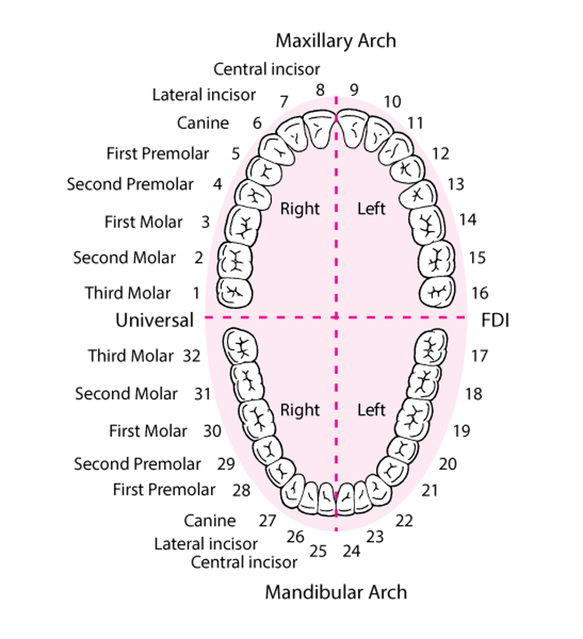
The teeth are categorized as incisors, canines, premolars, and molars and conventionally are numbered beginning with the maxillary right third molar (see figure Identifying the Teeth).
Identifying the Permanent Teeth
Although there are several tooth numbering systems, the one shown is the Universal Numbering System and is the one most commonly used in the United States. (Dental Education Hub: Teeth Names and Numbering Systems: You need to know. May 2020.) |

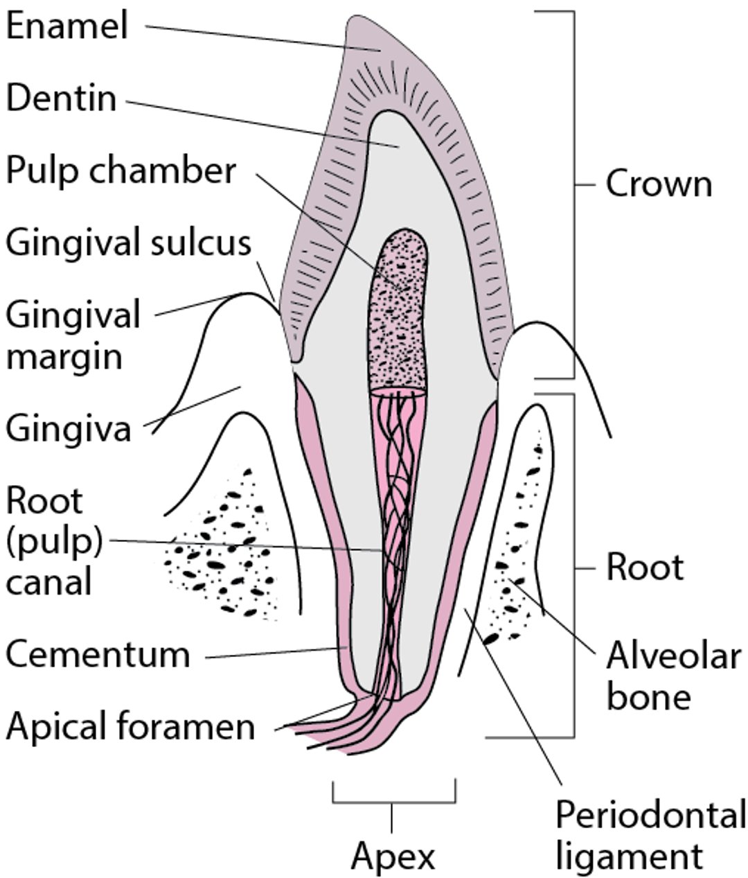
Each tooth has a crown and a root. The canines have the largest and strongest roots. An inner pulp contains blood vessels and nerves, surrounded by the hard but porous dentin, which is sensitive to touch and temperature changes. A very hard enamel coating covers the crown. The bone-like cementum is over the root, which, when healthy, is covered by gingiva (see figure Cross Section of a Canine Tooth).
Twenty deciduous teeth normally begin appearing at close to age 6 months and should all be in place by age 30 months (see table ). These teeth are followed by 32 permanent teeth that begin to appear by approximately age 6 years. The period from ages 6 to 11 is called the mixed dentition stage, in which both deciduous and permanent teeth are present. Timing of tooth eruption is one indicator of skeletal age and may identify growth restriction or establish age for forensic purposes.
(See also Introduction to the Dental Patient.)
Cross Section of a Canine Tooth

Supporting Tissues
The periodontium consists of the tissues that support the teeth—the gingiva, epithelial attachment, connective tissue attachment, periodontal ligament, and alveolar bone. The gingiva surrounds the teeth at the base of their crown. The alveolar ridges are trabecular bone containing sockets for the teeth. The mandible and maxilla support the alveolar ridges and house the teeth. Saliva from the salivary glands bathes and protects the teeth. The tongue directs food between the grinding surfaces and helps clean the teeth.
The maxilla is innervated by the maxillary nerve (V2), which is the second division of the trigeminal nerve (the 5th cranial nerve). The mandible is innervated by the mandibular nerve (V3), which is the third and most inferior division of the trigeminal nerve.

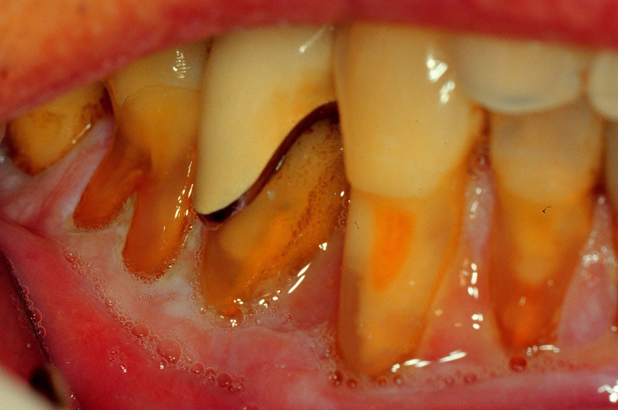
Recession of the gingivae exposes the root surfaces.
Image provided by Jonathan A. Ship, DMD.
In older adults and individuals with periodontal disease, gingival recession may expose the root surface adjacent to the crown, increasing the risk of root caries. If tooth destruction necessitates extraction, the mechanical stimulation necessary for maintaining bone integrity ceases. Consequently, atrophy of the alveolar ridge occurs following tooth loss.
Mouth
Keratinized epithelium is found on the facial surface of the lips, dorsum of the tongue, hard palate, and gingiva around the teeth. In health, the keratinized gingiva extends 1 to 5 mm apical to the crown of the tooth. Nonkeratinized mucosa covers the alveolar bone more distant from the crown, inside the lips and cheeks, on the sides and undersurface of the tongue, on the soft palate, and on the floor of the mouth. The junction between the skin and oral mucosa of the lips is demarcated by the vermilion border.
The buccal mucosa, including the vestibule and nonkeratinized alveolar mucosa, is usually smooth and moist. Innocuous entities in this region include the following:
Linea alba: appears as a thin white line, typically bilateral, on the level of the occlusal plane, resulting from cheek biting
Fordyce granules: aberrant sebaceous glands appearing as < 1 mm light yellow papules that also may occur on the lips
White sponge nevus: bilateral thick white folds over most of the buccal mucosa
Occasionally, pigmentation of the mucosa may arise from foreign material that is incorporated into the tissue. Most commonly, this occurs as a blue or black area adjacent to a dental amalgam filling. This is known as an amalgam tattoo. The orifices of the parotid (Stensen) ducts are opposite the maxillary first molar on the inside of each cheek and should not be mistaken for an abnormality. Recognizing these avoids needless biopsy and apprehension.


This photo shows a thin, white line in the buccal mucosa called linea alba. It occurs as a result of biting the cheek.
DR P. MARAZZI/SCIENCE PHOTO LIBRARY
The dorsal surface of the tongue is covered with numerous whitish elevations called the filiform papillae. Interspersed among them are isolated reddish prominences called the fungiform papillae, occurring mostly on the anterior part of the tongue. The circumvallate papillae, numbering 8 to 12, are considerably larger and lie posteriorly in a V pattern. The circumvallate papillae are surrounded by a trench. The foliate papillae appear as a series of parallel, slitlike folds on the lateral borders of the tongue, near the anterior pillars of the fauces. They vary in length and can easily be confused with malignant lesions, as may the foramen cecum (anatomical depression on posterior dorsal midline of tongue), median rhomboid glossitis (central papillary atrophy on dorsal tongue), and, rarely, a lingual thyroid nodule (ectopic thyroid tissue). Lingual tonsils, at the back of the tongue, are components of the Waldeyer ring and should not be mistaken for lesions. If an apparent abnormality is bilateral, it is almost always a normal variant.
Innervation is supplied by the lingual nerves (branches of the 5th cranial nerves) for general sensory innervation and the chorda tympani fibers (of the 7th cranial nerves) that innervate the taste buds of the anterior two thirds of the tongue. The glossopharyngeal nerves (9th cranial nerves) provide the sensations of touch and taste in the posterior third of the tongue behind the circumvallate papillae. The vagus nerve (10th cranial nerve) provides minor taste sensation to the region of the upper esophagus and epiglottis. The tongue has taste receptors for sweet, salty, sour, bitter, and umami (a savory taste triggered by natural glutamic acid and glutamates such as the flavoring agent monosodium glutamate). Although previously thought to be isolated to particular portions of the tongue, taste receptors are now known to be distributed over the surface of the tongue. The hypoglossal nerves (12th cranial nerves) control movement of the tongue.
Locating the Major Salivary Glands


The major salivary glands are the paired parotid, submandibular, and sublingual glands. Most oral mucosal surfaces contain many minor mucus-secreting salivary glands. Anteriorly and near the midline on each side of the floor of the mouth are the openings of the Wharton ducts, which drain the ipsilateral submandibular and sublingual glands. The parotid glands drain into the oral cavity via the Stensen ducts.