


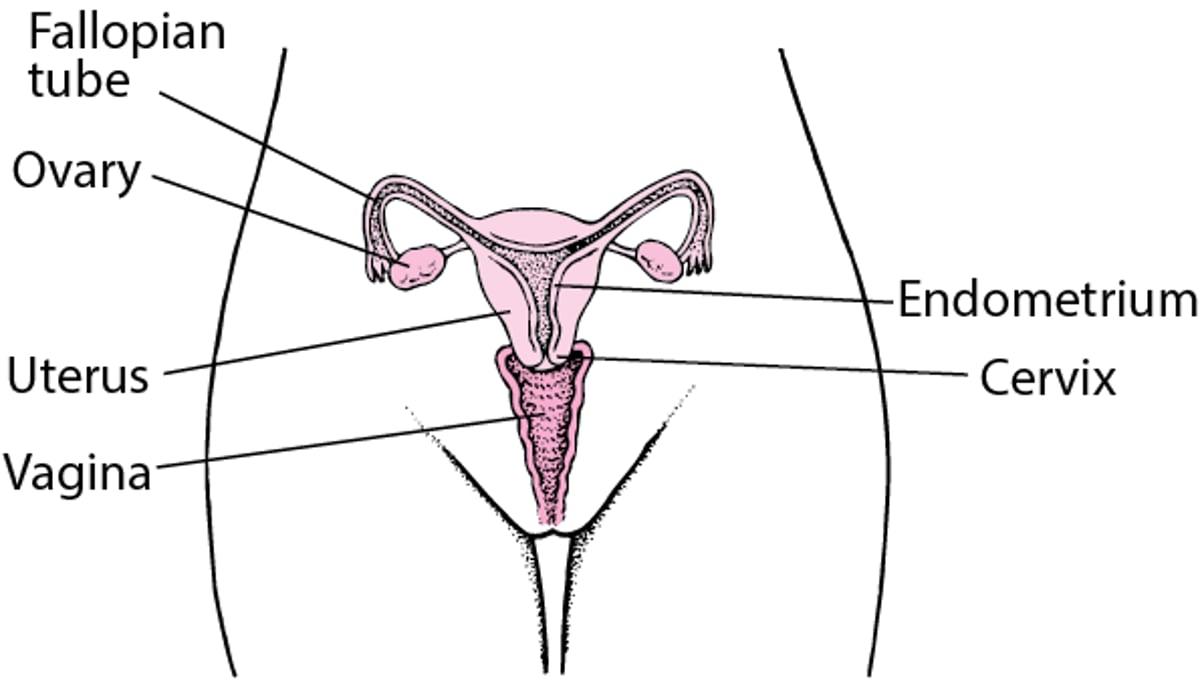
The female internal genital organs form a pathway (called the genital tract or reproductive tract). This pathway consists of the following:
Vagina (part of the birth canal), where sperm are deposited and from which a baby is born
Cervix (the lower part of the uterus), where sperm enter and which opens (dilates) when a pregnant woman is ready to gives birth
Uterus, the lining of which is shed during a menstrual period and where an embryo can develop into a fetus
Fallopian tubes (oviducts), where sperm can fertilize an egg after traveling through the cervix and uterus
Ovaries, which produce and release eggs as well as reproductive hormones
Sperm can travel up the tract, and eggs travel down the tract.
Internal Female Reproductive Anatomy

The hymen is a ring of tissue located just inside the opening of the vagina (see figure ). The hymen usually encircles the opening. Rarely, it completely covers the opening (called an imperforate hymen), making it impossible for menstrual blood to pass. In such cases, a procedure is done to open the hymen. The hymen may tear at the first attempt at sexual intercourse or it may not tear if it is soft and pliable. The hymen may also be torn during exercise or insertion of a tampon. Tearing usually causes slight bleeding. When the hymen tears, it may no longer be visible or it may form small tags of tissue around the vaginal opening.
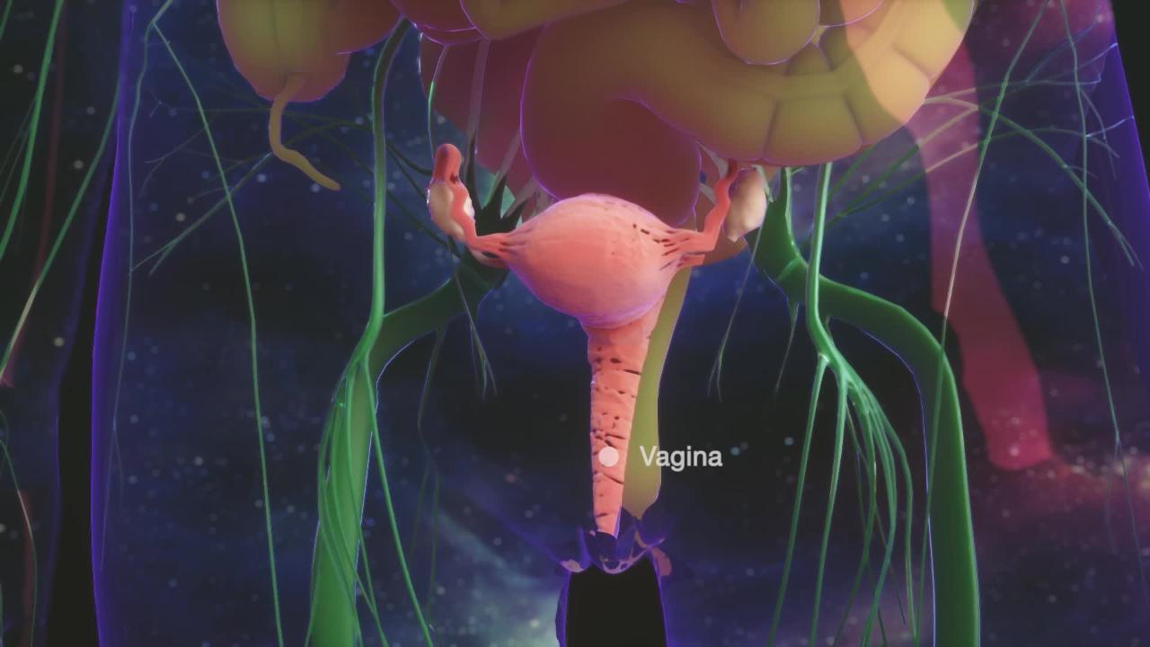
Vagina
The vagina is a soft, stretchable tube of muscle tissue about 4 to 5 inches long in an adult woman. It connects the external genital organs to the uterus. The upper part of the vagina is wider and surrounds the cervix (the lower part of the uterus). Some types of birth control devices (such as a diaphragm or vaginal ring) or other products related to contraception (such as spermicide gel) are inserted here.
The vagina has a central role in sexual activity and reproduction. It is the passageway for the following:
Sperm to the egg through the uterus and fallopian tubes
Menstrual bleeding or a baby to the outside

Because the vaginal tissue is soft, its walls can stretch for sexual intercourse, for childbirth, or for examination by a doctor (pelvic examination). After menopause, the vagina becomes less stretchy because estrogen levels decrease. This change can cause pain.
The vagina is lined with a type of tissue called a mucosal cells. These cells produce fluid that, along with secretions from the cervix, keep the vagina moist. A small amount of these fluids may pass to the outside as a small amount of clear or milky white vaginal discharge, which is normal. During a woman's reproductive years, the lining of the vagina has folds and wrinkles (called rugae). Before puberty and after menopause, the lining is smooth.
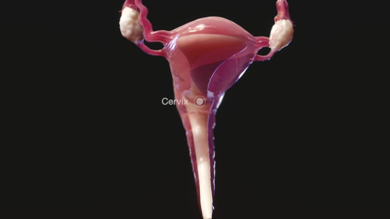
Uterus and cervix
The uterus is a thick-walled, muscular, pear-shaped organ located in the middle of the pelvis, behind the bladder, and in front of the rectum. The uterus is anchored in position by several ligaments. The main function of the uterus is to sustain a developing fetus.
The uterus consists of the following:
Cervix
Corpus (main body)
The cervix is the lower part of the uterus, which protrudes into the upper part of the vagina. During a pelvic examination, doctors can examine the cervix using a speculum (a metal or plastic instrument that spreads the walls of the vagina). Like the vagina, the cervix is lined with mucosal tissue.
Sperm can enter and menstrual blood can exit the uterus through a channel in the cervix (cervical canal). The cervical canal is usually narrow, but during labor, the canal dilates to let the baby through.
The cervix is usually a good barrier against bacteria. However, the bacteria that cause certain sexually transmitted infections can enter the uterus through the cervix during sexual intercourse.
Did You Know...
|
The channel through the cervix is lined with cells and glands that secrete mucus. This mucus is thick and impenetrable to sperm until just before ovulation. At ovulation, the mucus becomes clear and elastic (because the level of the hormone estrogen increases). As a result, sperm can swim through the mucus into the uterus to the fallopian tubes, where fertilization can take place.
Almost all pregnancies result from intercourse that occurs during the 3 days before ovulation. However, pregnancies sometimes result from intercourse that occurs up to 6 days before ovulation or during the 3 days after ovulation. For some women, the time between a menstrual period and ovulation varies from month to month. Consequently, pregnancy can occur at different times during a menstrual cycle.
The corpus of the uterus, which consists of muscle tissue, can stretch to accommodate a growing fetus. Its muscular walls contract during labor to push the baby out through the cervix and the vagina. During the reproductive years, the corpus is twice as long as the cervix. After menopause, the uterus and cervix are about the same length.
As part of a woman's reproductive cycle (which usually lasts about a month), the lining of the corpus (endometrium) thickens. If a woman does not become pregnant during that cycle, most of the endometrium is shed and bleeding occurs, resulting in the menstrual period.
How Many Eggs?
A baby girl is born with egg cells (oocytes) in her ovaries. By the fifth month of pregnancy, the ovaries of a female fetus contain about 7 million oocytes. Most of the oocytes gradually waste away, leaving about 1 to 2 million present at birth. No oocytes develop after birth. At puberty, only about 300,000—more than enough for a lifetime of fertility—remain. Only a small percentage of oocytes mature into eggs. The many thousands of oocytes that do not mature degenerate. Degeneration progresses more rapidly in the 10 to 15 years before menopause. All are gone by menopause. (Menopause is defined as 1 year after the last menstrual period.) Only about 400 eggs are released during a woman's reproductive life, usually one during each menstrual cycle. Until released, an egg remains dormant in its follicle—suspended in the middle of a cell division. Thus, the egg is one of the longest-lived cells in the body. Because a dormant egg cannot repair itself as cells usually do, the opportunity for damage increases as a woman ages. A chromosomal or genetic abnormality is thus more likely when a woman conceives a baby later in life. |
Fallopian tubes
The 2 fallopian tubes, which are about 4 to 5 inches (about 10 to 13 centimeters) long, extend from the upper edges of the uterus toward the ovaries. The tubes do not directly connect with the ovaries. Instead, the end of each tube flares into a funnel shape with fingerlike extensions (fimbriae). When an egg is released from an ovary, the fimbriae guide the egg into the opening of a fallopian tube.
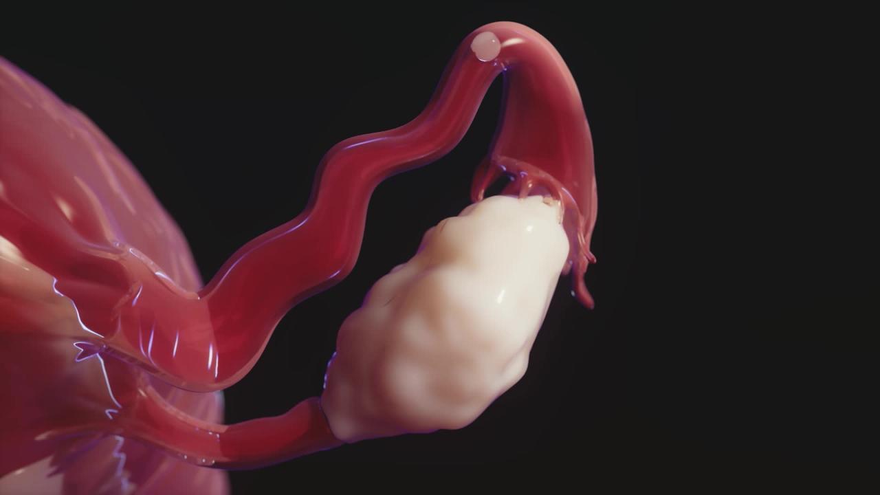
The fallopian tubes are lined with tiny hairlike projections (cilia). The cilia and the muscles in the tube's wall propel an egg downward through the tube to the uterus. The fallopian tube is the usual site of fertilization of the egg by the sperm. After fertilization, the fertilized egg enters the uterus and implants there.
Ovaries
The ovaries are usually pearl-colored, oblong, and about the size of a walnut. They are attached to the uterus by ligaments. In addition to producing female sex hormones (estrogen and progesterone) and some male sex hormones, the ovaries produce and release eggs. The developing egg cells (oocytes) are contained in fluid-filled cavities (follicles) in the wall of the ovaries. Each follicle contains one oocyte.


