

An evaluation of the foot includes a physical examination and sometimes arthrocentesis (see How To Do Metatarsophalangeal Joint Arthrocentesis).
(See also Evaluation of the Patient With Joint Symptoms.)
Physical Examination of the Foot
The patient is observed standing, for collapse of the longitudinal arch in pes planus (flat foot) and pes cavus (a high arch). The patient is observed from behind while standing on toes; contraction of the gastrocnemius, the degree of heel inversion, the height of the foot arch, and the degree of lateral symmetry are assessed.
The entire foot is inspected for discoloration, swelling, and lesions and for deformities such as hallux valgus (or bunion) of the great toe and hammer toe deformities of the other toes. Inspection between and underneath the toes may reveal lesions and sores unnoticed by the patient, particularly in diabetics (see figure ) and patients with peripheral neuropathy. Inspecting the patient’s shoes for abnormal or asymmetric wear patterns can help identify stresses that contribute to abnormalities such as osteoarthritis.
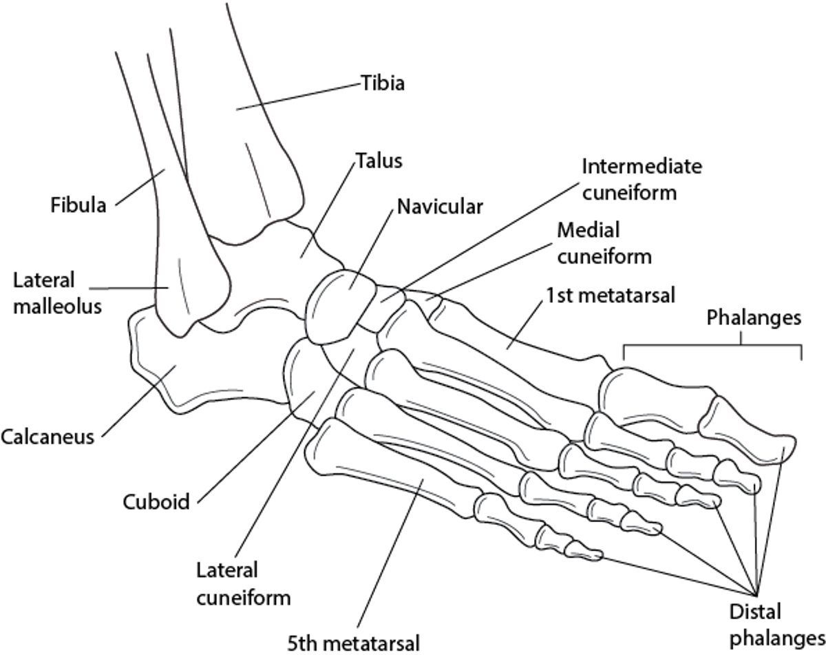
Bones of the Foot and Ankle

The foot is palpated gently for warmth and to detect subtle swelling. Comparison to the unaffected side is useful. Sensation to light touch is tested, at minimum, on the top of the first webbed space and the side of the foot. The dorsalis pedis pulse is palpated over the anterior foot, and the posterior tibial pulse is palpated behind the medial malleolus.


The foot is palpated for tenderness using one finger and beginning with light palpation to minimize patient anxiety. Palpation of the metatarsal bones and joints should include the 5th metatarsal head, a common site of fracture; the spaces between the metatarsal heads; and the tarsometatarsal joint. After injury, palpation of the tarsal bones should include the navicular, another common site of foot fracture. Patients with heel pain after injury are assessed for tenderness of the calcaneus by cupping the heel in one hand and squeezing.
Passive range of motion is tested for dorsiflexion, plantar flexion, eversion (while securing the heel), and inversion (by rotating the heel inward). Active range of motion is tested for dorsiflexion, plantar flexion, and eversion. Posterior tibialis function is tested by having the patient stand on one foot and attempt to rise onto the ball of the foot (single-heel-raise test).


