

Melanoma is a skin cancer that begins in the pigment-producing cells of the skin (melanocytes).
Melanomas can begin on normal skin or in existing moles.
They may be irregular, flat or raised brown patches of skin with spots of different colors or firm black or gray lumps.
To diagnose melanoma, doctors do a biopsy.
Melanomas are removed.
If they have spread, chemotherapy and radiation therapy are used, but cure is difficult.
Melanocytes are the pigment-producing cells in the skin that give skin its distinctive color. Sunlight stimulates melanocytes to produce more melanin (the pigment that darkens the skin) and increases the risk of melanoma.
In 2025, approximately 104,960 new cases of melanoma are estimated to occur in the United States, causing an estimated 8,430 deaths. Although melanoma accounts for less than 2% of all skin cancers diagnosed in the United States, it causes the most skin cancer deaths.
Melanoma usually begins on normal skin as a new, small, pigmented growth, most often on sun-exposed areas. About 1 in 3 melanomas develops in a preexisting mole. Melanoma may also occur around and inside the eyes, in the mouth, on the genitals and rectal areas, in the brain, and in the nail beds.
Melanoma readily spreads (metastasizes) to distant parts of the body, where it continues to grow and destroy tissue.
The two most common types of melanoma are:
Superficial spreading melanoma: This type accounts for 70% of melanomas and occurs most commonly on women’s legs and men’s torsos. The tumor cells commonly have mutations in the BRAF gene.
Lentigo maligna melanoma: This type accounts for 15% % of melanomas, and usually occurs on the face or other areas of chronic sun exposure.
Risk Factors for Melanoma
Risk factors for melanoma include the following:
Sun exposure (mainly repeated blistering sunburns)
Repeated tanning with ultraviolet A (UVA) or medical treatment with psoralen plus ultraviolet A (PUVA)
Personal history of skin cancer (another melanoma or another type of skin cancer)
Family history of breast, ovarian, or pancreatic cancer
Family members with melanoma
Light skin, freckling
Large numbers of pigmented moles (>50) or presence of atypical moles (especially more than 5)
A weakened immune system
A large congenital melanocytic nevus (giant congenital nevus)
Mutations in certain genes, such as BRCA2. BRAF, CDKN2A (p16), BAP1, and CHEK2
Advanced age
People who have had melanoma are at increased risk of developing a new melanoma.

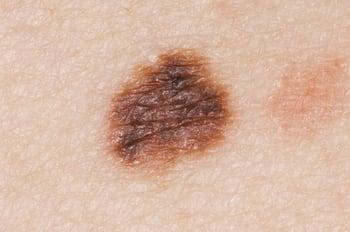
Features of this atypical mole include irregular borders and variable colors.
Melanoma is less common among people who have dark skin. When melanoma does develop in people who have dark skin, it often develops in the nail beds and on the palms and soles.
Melanomas are very rare in childhood. However, congenital melanocytic nevus is a dark-colored patch of skin, like a mole or a birthmark, that is present at birth. When large in size, for example, more than about 8 inches (about 20 centimeters), congenital melanocytic nevus is a risk factor for malignant melanoma.

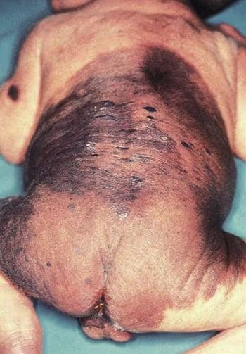
Congenital melanocytic nevus (giant congenital nevus), when large, is a risk factor for malignant melanoma. The nevus in this image is more than about 8 inches (about 20 centimeters), has an irregular border, and has several different colors.
Although melanomas occur during pregnancy, pregnancy does not increase the likelihood that a mole will become a melanoma. Moles frequently change in size and darken during pregnancy.
All people should know the ABCDEs of melanoma so they can check their moles for any malignant (cancerous) changes.
Symptoms of Melanoma
Melanomas can vary in appearance. Some are flat, irregular brown patches containing small black spots. Others are raised brown patches with red, white, black, or blue spots. Sometimes melanoma appears as a firm red, black, or gray lump.
Two to 8% of melanomas produce no pigment. These so-called amelanotic melanomas may be pink, red, or slightly light-brown and may look like noncancerous growths or a form of nonmelanoma skin cancer.


This photo highlights the irregular border of superficial spreading melanoma.
This photo highlights the irregular border of superficial spreading melanoma.
Image provided by Gregory L. Wells, MD.


This raised growth is characteristic of nodular melanoma, which usually appears gray to black.
This raised growth is characteristic of nodular melanoma, which usually appears gray to black.
Photo courtesy of Gregory L. Wells, MD.


This photo shows a nodular melanoma. It has dark, reddish brown to black discoloration; a raised texture; and shapeless, irregular borders.
This photo shows a nodular melanoma. It has dark, reddish brown to black discoloration; a raised texture; and shapeless
CDC/ Carl Washington, M.D., Emory Univ. School of Medicine; Mona Saraiya, MD, MPH


This photo shows an asymmetric melanoma. It is asymmetric, and the borders are red and dark brown. It is large in size.
This photo shows an asymmetric melanoma. It is asymmetric, and the borders are red and dark brown. It is large in size.
National Cancer Institute (NCI); www.cancer.gov


This photo shows an asymmetric melanoma. It ranges from pinkish red to dark brown/black in color, and the borders are very irregular.
This photo shows an asymmetric melanoma. It ranges from pinkish red to dark brown/black in color, and the borders are v
National Cancer Institute (NCI); www.cancer.gov


This photo shows a darkly pigmented, raised growth that is a superficial spreading melanoma (left). The raised, smooth, red bump (nodule) also has less pigment and is considered an amelanotic melanoma (right).
This photo shows a darkly pigmented, raised growth that is a superficial spreading melanoma (left). The raised, smooth,
National Cancer Institute (NCI); www.cancer.gov


Sometimes melanomas may not have any pigment (that is, no brown, black, or blue discoloration). This photo shows an amelanotic melanoma.
Sometimes melanomas may not have any pigment (that is, no brown, black, or blue discoloration). This photo shows an ame
DR P. MARAZZI/SCIENCE PHOTO LIBRARY
This photo highlights the irregular border of superficial spreading melanoma.
This photo highlights the irregular border of superficial spreading melanoma.
Image provided by Gregory L. Wells, MD.
This raised growth is characteristic of nodular melanoma, which usually appears gray to black.
This raised growth is characteristic of nodular melanoma, which usually appears gray to black.
Photo courtesy of Gregory L. Wells, MD.
This photo shows a nodular melanoma. It has dark, reddish brown to black discoloration; a raised texture; and shapeless, irregular borders.
This photo shows a nodular melanoma. It has dark, reddish brown to black discoloration; a raised texture; and shapeless
CDC/ Carl Washington, M.D., Emory Univ. School of Medicine; Mona Saraiya, MD, MPH
This photo shows an asymmetric melanoma. It is asymmetric, and the borders are red and dark brown. It is large in size.
This photo shows an asymmetric melanoma. It is asymmetric, and the borders are red and dark brown. It is large in size.
National Cancer Institute (NCI); www.cancer.gov
This photo shows an asymmetric melanoma. It ranges from pinkish red to dark brown/black in color, and the borders are very irregular.
This photo shows an asymmetric melanoma. It ranges from pinkish red to dark brown/black in color, and the borders are v
National Cancer Institute (NCI); www.cancer.gov
This photo shows a darkly pigmented, raised growth that is a superficial spreading melanoma (left). The raised, smooth, red bump (nodule) also has less pigment and is considered an amelanotic melanoma (right).
This photo shows a darkly pigmented, raised growth that is a superficial spreading melanoma (left). The raised, smooth,
National Cancer Institute (NCI); www.cancer.gov
Sometimes melanomas may not have any pigment (that is, no brown, black, or blue discoloration). This photo shows an amelanotic melanoma.
Sometimes melanomas may not have any pigment (that is, no brown, black, or blue discoloration). This photo shows an ame
DR P. MARAZZI/SCIENCE PHOTO LIBRARY
Diagnosis of Melanoma
Biopsy
A new mole or changes in a mole—such as enlargement (especially with an irregular border), darkening, inflammation, spotty color changes, bleeding, itching, tenderness, and pain—are warning signs of possible melanoma and so are the ABCDEs of melanoma. There are additional tools that doctors may use to help distinguish an atypical mole from a melanoma. These tools include polarized light and dermoscopy, which help to better evaluate the growths. Visualizing the mole under a dermoscope may help doctors identify cancerous moles. If these or other findings lead doctors to suspect melanoma, they do a biopsy.
For the biopsy, doctors remove the entire growth if it is small or only part of it if it is large. Doctors look at many different aspects of a biopsied tissue to identify (ie, determine the type of melanoma) and stage the cancer. They then examine the sample under a microscope to determine whether the growth is a melanoma and, if so, whether all the cancer has been removed. If the biopsy shows that the growth is a melanoma and the growth has not been completely removed, it is then completely removed. A growth that is a melanoma is often tested for genetic mutations because results may further help doctors determine treatment.
Most darkly pigmented growths that are removed for biopsy are not melanoma but, rather, simple moles. Nonetheless, removing even many harmless moles is preferable to allowing a single cancer to grow. Some growths are neither simple moles nor melanomas, but something in between. These growths, called atypical moles (dysplastic nevi), sometimes turn into melanoma later.
Sometimes, additional biopsies such as a sentinel lymph node biopsy may be done if spread to the lymph nodes is suspected. These types of biopsies are recommended in patients with deeper or thicker melanomas. Sentinel lymph node biopsies and can also be considered for thinner melanomas with higher risk features, particularly in younger patients or those without other medical issues.
Treatment of Melanoma
Removal of the tumors
Possibly imiquimod, cryotherapy, or radiation therapyPossibly imiquimod, cryotherapy, or radiation therapy
For tumors that have spread, immunotherapy, targeted therapy, chemotherapy, or radiation therapy
Doctors treat melanomas by cutting them out (sometimes using Mohs microscopically controlled surgery), taking a border of almost ½ inch (1 centimeter) or more of skin around the tumor.
For people who have the most shallow melanomas (that is, melanomas that have not invaded past the epidermis—called melanoma in situ) and who cannot have surgery (for example, because their health is too poor) or choose not to (for example, because their melanomas are in cosmetically important areas), doctors may treat with imiquimod cream or may use extreme cold (cryosurgery) to destroy the melanomas.For people who have the most shallow melanomas (that is, melanomas that have not invaded past the epidermis—called melanoma in situ) and who cannot have surgery (for example, because their health is too poor) or choose not to (for example, because their melanomas are in cosmetically important areas), doctors may treat with imiquimod cream or may use extreme cold (cryosurgery) to destroy the melanomas.
Did You Know...
|
Melanoma that has spread
If melanoma has spread to distant areas (metastasized), surgery is generally not an option, but sometimes localized areas of cancer (for example, the affected lymph nodes) may be surgically removed.
The immunotherapies pembrolizumab and nivolumab are used to help the body's immune system destroy the cancer. These medications are called PD-1 inhibitors because they block the action of a protein on the surface of the cancer cell called programmed cell death protein 1. This protein protects the cancer cell from the effects of the immune system. When PD-1 inhibitors block the protein, the immune system is able to attack the cancer cell and kill it. PD-1 inhibitors are proving to be very effective treatments for metastatic melanoma. Ipilimumab is another immunotherapy that improves survival by activating certain white blood cells to attack cancer cells. The combination of pembrolizumab and nivolumab are used to help the body's immune system destroy the cancer. These medications are called PD-1 inhibitors because they block the action of a protein on the surface of the cancer cell called programmed cell death protein 1. This protein protects the cancer cell from the effects of the immune system. When PD-1 inhibitors block the protein, the immune system is able to attack the cancer cell and kill it. PD-1 inhibitors are proving to be very effective treatments for metastatic melanoma. Ipilimumab is another immunotherapy that improves survival by activating certain white blood cells to attack cancer cells. The combination ofnivolumab and ipilimumab is often the preferred treatment. Nivolumab can also be used in combination with another immunotherapy called relatlimab, which has been found to cause fewer side effects that some other combinations while still being effective. Sometimes PD-1 inhibitors are used before surgery to reduce the chances of the cancer returning.
Targeted therapy consists of medications that attack a cancer cell's innate biologic mechanisms. In targeted therapy, medications attack specific parts of cells that are present only in the cancerous cells with specific gene mutations. Use of these medications has improved cancer survival in some people. One class of medications targets cells with a BRAF gene mutation. These medications include dabrafenib, encorafenib, and vemurafenib. For people who cannot be treated with immunotherapy, a combination of targeted therapies can be used.gene mutation. These medications include dabrafenib, encorafenib, and vemurafenib. For people who cannot be treated with immunotherapy, a combination of targeted therapies can be used.
Chemotherapy medications, such as dacarbazine and temozolomide, have not been proved to prolong survival, but they are sometimes given to treat melanomas that have spread in people who do not have other treatment options. medications, such as dacarbazine and temozolomide, have not been proved to prolong survival, but they are sometimes given to treat melanomas that have spread in people who do not have other treatment options.
Radiation therapy may be used in people when complete removal of a melanoma is not possible because of its location, when it recurs in an area where it had originally been removed, and when it has spread to the brain.
Other treatments are being investigated, such as medications (eg, T-VEC) and vaccines that stimulate the body to attack the melanoma cells.
Prognosis for Melanoma
Melanoma may spread rapidly and cause death within months of diagnosis. The less a melanoma has grown deeper into the skin, the greater the chance that surgery will cure it. Almost 100% of the earliest, most shallow melanomas are cured by surgery. However, melanomas that have grown deeper than about 1/32 inch (about 1 millimeter) into the skin have a higher risk of metastasizing to the lymph nodes and blood vessels.
The 5-year survival rate is approximately 95%. However, this drops dramatically if cancer has spread. In general, the course of the disease varies greatly and depends in part on the strength of the body’s immune defenses. Some people survive in apparent good health for several years despite the spread of the melanoma.
Prevention of Melanoma
Because melanoma is caused by long-term sun exposure, people can help prevent this cancer by doing the following, starting in early childhood:
Avoiding the sun: For example, seeking shade, minimizing outdoor activities between 10 AM and 4 PM (when the sun’s rays are strongest), and avoiding sunbathing and the use of tanning beds (particularly adolescents and young adults)
Wearing protective clothing: For example, long-sleeved shirts, pants, and broad-brimmed hats
Using sunscreen: At least sun protection factor (SPF) 30 with UVA and UVB protection used as directed and reapplied every 2 hours and after swimming or sweating but not used to prolong sun exposure
Doctors do not know with certainty whether these measures decrease the chances of people developing or dying of melanoma. However, using tanning beds, particularly by young people, does seem to increase the risk of melanoma.
Anyone who has had a melanoma is at risk of developing other melanomas. Therefore, such people need regular skin examinations.
People who have many moles should have a total body skin examination at least once a year. People can be taught to examine themselves to detect changes in existing moles and to recognize features suggesting melanoma. In people without risk factors, doctors do not know whether routine yearly skin examinations reduce the number of deaths from melanoma.
More Information
The following English-language resources may be useful. Please note that The Manual is not responsible for the content of these resources.
Melanoma Research Foundation: Information about various types of melanoma, current research, and clinical trials
American Cancer Society: Melanoma Skin Cancer: Information about melanoma, including detection, prevention, treatment options, and other resources
The Skin Cancer Foundation: Melanoma Overview: Information about melanoma, including detection, prevention, treatment options, and other resources


