

Contagious ecthyma and milker's nodules are two viral skin diseases that can rarely be transmitted from animals to humans.
Contagious ecthyma
Contagious ecthyma (contagious pustular dermatitis), also called orf, is caused by the orf virus, a parapoxvirus that infects ruminants (most often sheep and goats). Farmers, veterinarians, zoo caretakers, and others with direct animal contact are at risk.
Contagious ecthyma is different from ecthyma gangrenosum, which is a rare cutaneous lesion that can occur with Pseudomonas aeruginosa infection in immunocompromised patients.
The cutaneous findings pass through 6 stages that together last approximately 1 week:
Stage 1 (papular): A single red edematous papule on a finger (most commonly right index finder)
Stage 2 (target): A larger nodule with a red center surrounded by a white ring with a red periphery
Stage 3 (acute): A rapidly growing, infected-looking tumor
Stage 4 (regenerative): A nodule with black dots covered with a thin transparent crust
Stage 5 (papillomatous): A nodule with a surface studded with small projections
Stage 6 (regressive): A flattened nodule with a thick crust

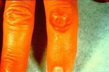
Contagious ecthyma is a focal skin lesion caused by orf virus infection, usually occurring on a finger and transmitted to animal workers from sheep or goats. The lesion passes through 6 stages; this image shows the 2nd stage (target), characterized by a large nodule with an erythematous center surrounded by a white ring and an erythematous periphery.
Patients can develop regional adenopathy, lymphangitis, and fever.
Diagnosis of contagious ecthyma is by history of contact; differential diagnosis is extensive depending on the stage of the lesion. Acute lesions must be differentiated from milker’s nodules, Mycobacterium marinum infection, and other bacterial infections; regressed lesions must be differentiated from cutaneous tumors, such as Bowen disease or squamous cell carcinoma.
Lesions heal spontaneously; no treatment is necessary.
Milker’s nodules
These nodules are caused by paravaccinia virus, a parapoxvirus that causes udder lesions in cows. Infection requires direct contact and causes macules that progress to papules, vesicles, and nodules. This infection has 6 stages, which are similar to those of contagious ecthyma. Fever and lymphadenopathy are uncommon.
Diagnosis of milker's nodules is by history of contact and cutaneous findings. Differential diagnosis varies depending on morphology but can include primary inoculation tuberculosis (a chancre that can develop at the site of tuberculosis inoculation), sporotrichosis, anthrax, and tularemia.
Lesions heal spontaneously; no treatment is necessary.

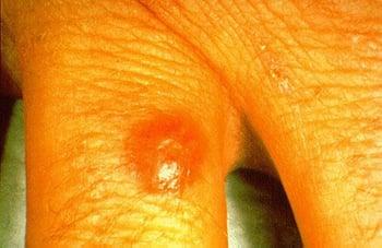
Milker’s nodule is a focal skin lesion caused by paravaccinia virus, a parapoxvirus that causes udder lesions in cows. The lesion passes through 6 stages; this image shows the stage in which a single erythematous edematous papule develops from an initial macule.
More Information
The following English-language resources may be useful.
MSD Veterinary Manual: Overview of Contagious Ecthyma
MSD Veterinary Manual: Pseudocowpox in Cattle


