

Infantile hemangiomas are raised, red or purplish, hyperplastic vascular lesions appearing in the first year of life. Most spontaneously involute; those obstructing vision, the airway, or other structures require treatment. Ideal treatment varies based on many patient-specific factors.
Infantile hemangioma is the most common tumor of infancy, affecting 5 to 10% of infants by age 1 year (1). Infantile hemangioma occurs almost always within the first several weeks of life; occasionally, deeper lesions may not be apparent until a few months after birth. Size and vascularity increase rapidly, reaching 80% of their final size by 3 months of age; the majority of hemangiomas will have completed growth by 5 months of age (2). Most deep hemangiomas grow between 0.5 and 5 cm across, although sometimes they grow much larger.
Infantile hemangiomas can be classified by their general appearance (superficial, deep, or cavernous) or by other descriptive terms (eg, strawberry hemangioma). However, because all of these lesions share a common pathophysiology and natural history, the inclusive term infantile hemangioma is preferred.
References
1. Tiemann L, Hein S: Infantile hemangioma: A review of current pharmacotherapy treatment and practice pearls. J Pediatr Pharmacol Ther 25(7):586-599, 2020. doi: 10.5863/1551-6776-25.7.586
2. Krowchuk DP, Frieden IJ, Mancini AJ, et al. Clinical Practice Guideline for the Management of Infantile Hemangiomas. Pediatrics. 2019;143(1):e20183475. doi:10.1542/peds.2018-3475
Symptoms and Signs of Infantile Hemangiomas
The appearance of skin lesions varies by depth: superficial ones are bright red, and deeper ones are bluish. Minor trauma may lead to bleeding or even painful ulceration.
Infantile hemangiomas in certain locations can interfere with physiologic functions. For instance, lesions on the face or oropharynx may interfere with vision or obstruct the airway; those near the urethral meatus or anus may interfere with elimination. A periocular hemangioma in an infant is considered an emergency and should be attended to promptly to avoid permanent visual defects. Lumbosacral hemangiomas may be a sign of underlying neurologic or genitourinary anomalies (1, 2).

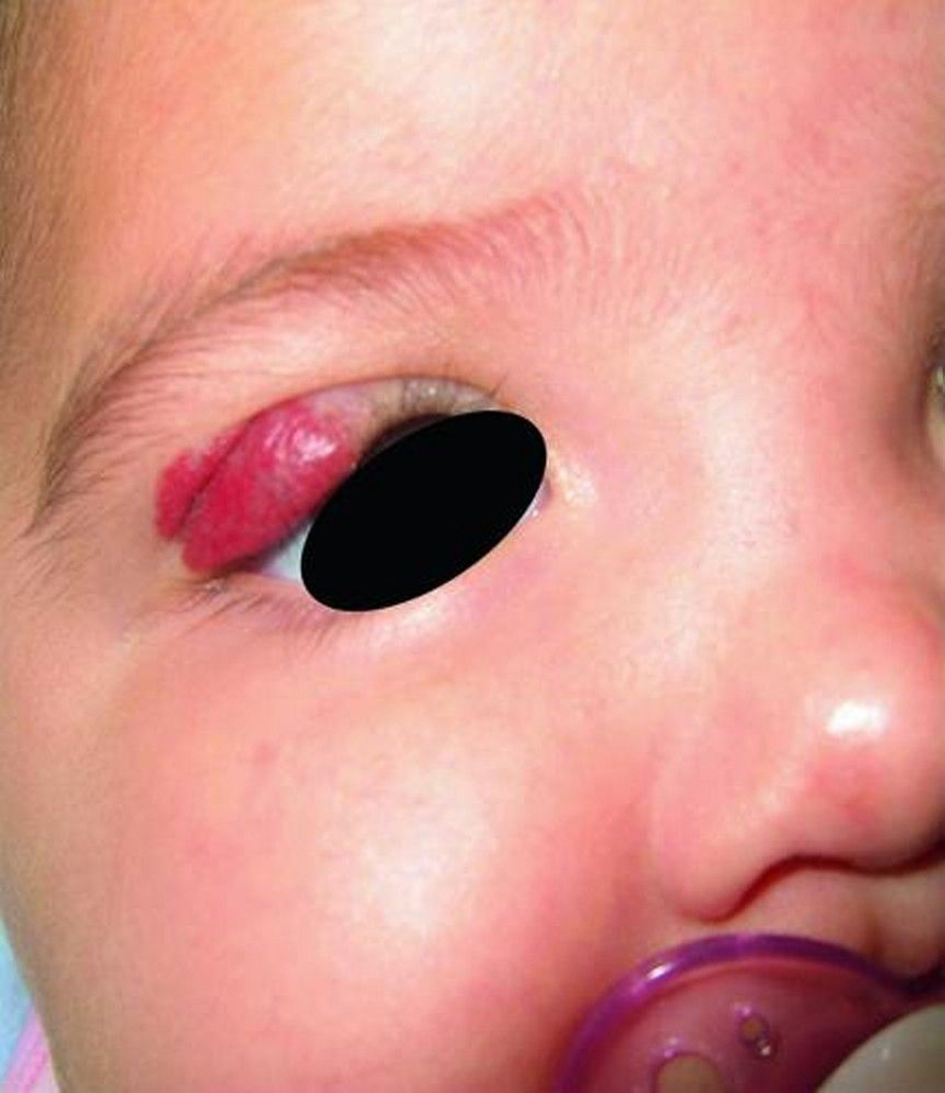
Periocular hemangiomas are hemangiomas surrounding the eye.
© Springer Science+Business Media
Lesions slowly involute starting at 12 to 18 months, decreasing in size and vascularity. Many infantile hemangiomas regress spontaneously within the first 3 to 5 years of life. Involuted lesions commonly have a yellowish or telangiectatic color and a wrinkled or lax fibrofatty texture. Residual changes are almost always proportional to the lesion’s maximal size and vascularity.
Symptoms and signs references
1.Girard C, Bigorre M, Guillot B, Bessis D. PELVIS Syndrome. Arch Dermatol. 2006;142(7):884-888. doi:10.1001/archderm.142.7.884
2. Iacobas I, Burrows PE, Frieden IJ, et al. LUMBAR: association between cutaneous infantile hemangiomas of the lower body and regional congenital anomalies. J Pediatr. 2010;157(5):795-801.e8017. doi:10.1016/j.jpeds.2010.05.027
Diagnosis of Infantile Hemangiomas
Primarily physical examination
The diagnosis of infantile hemangiomas is clinical; the extent can be evaluated by MRI if lesions appear to encroach on vital structures.
Treatment of Infantile Hemangiomas
For superficial or uncomplicated lesions that require treatment, possibly topical or intralesional glucocorticoids or topical beta-blockers (1)
For complicated or high-risk lesions requiring treatment, oral propranolol (For complicated or high-risk lesions requiring treatment, oral propranolol (2)
General wound care for ulcerated lesions
Usually avoidance of surgery
Because most lesions resolve spontaneously, observation is usually indicated before initiating treatment.
There is no universal infantile hemangioma treatment recommendation (3). The American Academy of Pediatrics recommends that treatment be individualized based on location, size, and severity of lesions. Treatment should be considered for complicated or high-risk lesions, ie, those that:
Threaten life
Threaten function (eg, vision)
Involve large areas of the face
Are distributed over the beard area
Are ulcerated
Are multiple
Are lumbosacral
Topical treatments and wound care are useful for ulcerated lesions and help prevent scarring, bleeding, and pain. Compresses, topical mupirocin or metronidazole, barrier dressings (generally polyurethane film dressing or petrolatum-impregnated gauze), or barrier creams may be used to prevent infection and/or reduce colonization.Topical treatments and wound care are useful for ulcerated lesions and help prevent scarring, bleeding, and pain. Compresses, topical mupirocin or metronidazole, barrier dressings (generally polyurethane film dressing or petrolatum-impregnated gauze), or barrier creams may be used to prevent infection and/or reduce colonization.
Unless complications are life threatening or vital organs are compromised, surgical excision or other destructive procedures should be avoided because they frequently cause more scarring than occurs with spontaneous involution. To help parents better accept nonintervention, the physician can review the natural history (photographic examples are helpful), conduct serial photography of the lesion to document involution, and provide reassurance for any parental concerns.
Treatment references
1. Boos MD, Castelo-Soccio: Experience with topical timolol maleate for the treatment of ulcerated infantile hemangiomas (IH). : Experience with topical timolol maleate for the treatment of ulcerated infantile hemangiomas (IH).J Am Acad Dermatol 74(3):567-570, 2016. doi: 10.1016/j.jaad.2015.10.021
2. Hogeling M, Adams S, Wargon O: A randomized controlled trial of propranolol for infantile hemangiomas. : A randomized controlled trial of propranolol for infantile hemangiomas.Pediatrics 128(2):e259-266, 2011. doi: 10.1542/peds.2010-0029
3. Krowchuk DP, Frieden IJ, Mancini AJ, et al. Clinical Practice Guideline for the Management of Infantile Hemangiomas. Pediatrics. 2019;143(1):e20183475. doi:10.1542/peds.2018-3475
Key Points
Infantile hemangiomas affect 5 to 10% of infants by age 1 year.
Lesions slowly involute starting at 12 to 18 months.
Use topical treatments and wound care for ulcerated lesions and to help prevent scarring, bleeding, and pain.
Unless complications are life threatening or vital organs are compromised, avoid surgery.
Drug Information for the Topic


