

Orbital tumors can be benign or malignant and arise primarily within the orbit or secondarily from an adjacent source, such as the eyelid, paranasal sinus, or intracranial compartment. Orbital tumors can also be metastatic from distant sites.
Some types of orbital tumors cause proptosis and displacement of the globe superiorly or inferiorly, in a direction opposite the tumor. Pain, diplopia, and vision loss may also be present. The diagnosis of orbital tumors is suspected based on the history, examination, and neuroimaging (CT, MRI, or both), but confirmation often ultimately requires a biopsy. Causes and treatment vary by age group.
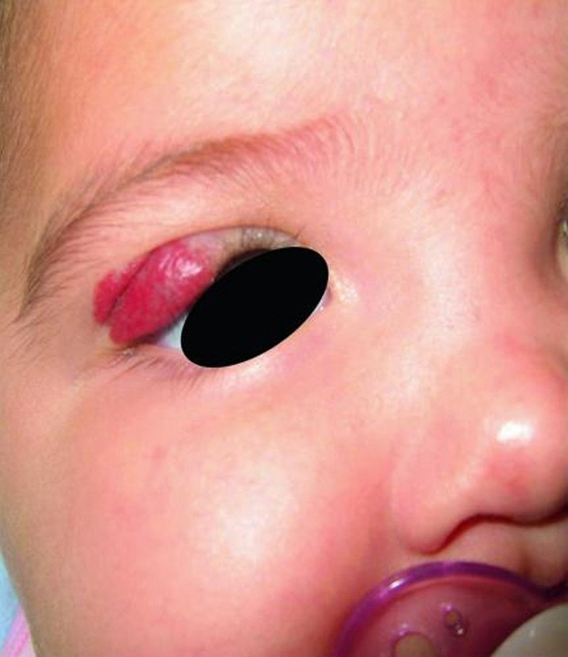
Children
Benign pediatric tumors are most commonly dermoid cysts and vascular lesions such as infantile hemangioma and lymphatic malformations (1). Treatment of dermoid cysts is excision. Infantile hemangiomas tend to spontaneously involute and therefore usually do not need treatment; however, especially when located on the upper eyelid, they may affect vision and require treatment with systemic beta-blockers (2). Small lymphatic malformations that do not cause symptoms may be followed clinically. For symptomatic or larger lymphatic malformations, options include surgical debulking, intralesional sclerotherapy, and sirolimus (). Small lymphatic malformations that do not cause symptoms may be followed clinically. For symptomatic or larger lymphatic malformations, options include surgical debulking, intralesional sclerotherapy, and sirolimus (3).

Periocular hemangiomas are hemangiomas surrounding the eye.
© Springer Science+Business Media
Malignant pediatric tumors are most commonly rhabdomyosarcoma and metastatic lesions related to leukemia or neuroblastoma. Management of rhabdomyosarcoma typically involves biopsy for diagnosis, followed by chemotherapy and orbital radiation therapy (4). Leukemic disease is usually managed by orbital radiation therapy, chemotherapy, or both.
Adults
Benign tumors in adults are most commonly meningiomas, mucoceles, and cavernous venous malformations (5). Pleomorphic adenomas of the lacrimal gland are less common. When symptomatic, sphenoid wing meningiomas are treated with debulking via craniotomy, sometimes followed by a course of radiation therapy. Because meningioma cells infiltrate bone of the skull base, complete resection usually is not possible. Mucoceles most commonly arise from the ethmoid or frontal sinus and are treated by draining them into the nose. Cavernous venous malformations (the most common benign orbital tumor) and lacrimal gland pleomorphic adenomas are excised.


In this image, the axial (top) and coronal (bottom) CT scans show a well-demarcated intraconal mass behind the left globe. The mass is a cavernous venous malformation.
Malignant tumors in adults are most commonly lymphoma, squamous cell carcinoma, and metastatic disease. Less commonly, the tumor is an adenoid cystic carcinoma of the lacrimal gland, which is an aggressive tumor.
Lymphomas involving the orbit are the most frequent malignant orbital tumor and are typically B-cell and characteristically low grade (usually a MALT lymphoma [mucosa-associated lymphoid tissue], also known as extranodal marginal zone B-cell lymphoma) (6). Lymphomas can be bilateral and simultaneous and can be part of a systemic process or exist in the orbit in isolation. Many orbital lymphomas have minimal symptoms and findings despite impressive radiologic findings. Radiation therapy effectively treats orbital lymphomas with few adverse effects. Treatment with monoclonal antibodies (eg, rituximab) against a surface receptor (eg, CD20) on the lymphocyte is also effective and should be considered in addition to or instead of radiation therapy, particularly if lymphoma is systemic. ). Lymphomas can be bilateral and simultaneous and can be part of a systemic process or exist in the orbit in isolation. Many orbital lymphomas have minimal symptoms and findings despite impressive radiologic findings. Radiation therapy effectively treats orbital lymphomas with few adverse effects. Treatment with monoclonal antibodies (eg, rituximab) against a surface receptor (eg, CD20) on the lymphocyte is also effective and should be considered in addition to or instead of radiation therapy, particularly if lymphoma is systemic.


In this image, the first coronal CT scan (left) shows a homogeneous mass conforming to the globe (red arrow). The second coronal CT scan (center) shows a well-demarcated mass in the right lacrimal gland fossa (blue arrow). The axial CT scan (right) shows a homogenous lacrimal fossa mass conforming to the right globe (yellow arrow).
Most squamous cell carcinomas arise from the adjacent paranasal sinuses or skin. Surgery, radiation therapy, or both form the backbone of therapy. PD-1 inhibitors are also effective in the treatment of cutaneous squamous cell carcinomas that have spread into the orbit (7).
Metastatic disease is usually treated with radiation therapy. Metastatic disease involving the orbit is usually an unfavorable prognostic sign; neuroendocrine tumors are a notable exception (8).
Lacrimal gland adenoid cystic carcinoma is treated with surgery and then usually with radiation therapy (sometimes proton beam therapy) or by a protocol using intra-arterial chemotherapy with radiation therapy and surgery (9, 10).
References
1. Shields JA, Bakewell B, Augsburger JJ. Space-occupying orbital masses in children. A review of 250 consecutive biopsies. Ophthalmology. 1986;93(3):379-384. doi: 10.1016/s0161-6420(86)33731-x
2. Hutchinson AK, Kraker RT, Pineles SL, et al. The Use of β-Blockers for the Treatment of Periocular Hemangiomas in Infants: A Report by the American Academy of Ophthalmology. Ophthalmology. 2019;126(1):146-155. doi:10.1016/j.ophtha.2018.07.023
3. Shoji MK, Shishido S, Freitag SK. The use of sirolimus for treatment of orbital lymphatic malformations: A systematic review. Ophthalmic Plast Reconstr Surg. 2020;36(3):215-221. doi: 10.1097/IOP.0000000000001518
4. Agarwal A, Vempuluru VS, Kaliki S. Primary ocular, adnexal, and orbital rhabdomyosarcoma: A review. Surv Ophthalmol. 2025;70(5):868-881. doi:10.1016/j.survophthal.2025.03.009
5. Bonavolontà G, Strianese D, Grassi P, et al. An analysis of 2,480 space-occupying lesions of the orbit from 1976 to 2011. Ophthalmic Plast Reconstr Surg. 2013;29(2):79-86. doi: 10.1097/IOP.0b013e31827a7622
6. Olsen TG, Heegaard S. Orbital lymphoma. Surv Ophthalmol. 2019;64(1):45-66. doi:10.1016/j.survophthal.2018.08.002
7. Rana K, Beecher M, Tong JY, et al. Immunotherapy for orbital squamous cell carcinoma. Orbit. 2025;44(5):504-510. doi:10.1080/01676830.2025.2469305
8. Allen RC. Orbital Metastases: When to Suspect? When to biopsy? Middle East Afr J Ophthalmol. 2018;25(2):60-64. doi:10.4103/meajo.MEAJO_93_18
9. Tse DT, Benedetto PW, Tse BC, Feuer WJ. Neoadjuvant Intra-Arterial Cytoreductive Chemotherapy for Lacrimal Gland Adenoid Cystic Carcinoma: A Long-Term Follow-up Study of a Trimodal Strategy. Am J Ophthalmol. 2022;240:239-251. doi:10.1016/j.ajo.2022.03.027
10. Woo KI, Kim YD, Sa HS, Esmaeli B. Current treatment of lacrimal gland carcinoma. Curr Opin Ophthalmol. 2016;27(5):449-456. doi:10.1097/ICU.0000000000000301


