

Interstitial keratitis is chronic, nonulcerative inflammation of the mid-stroma (the middle layers of the cornea) that is sometimes associated with uveitis. The cause is usually infectious. Symptoms are photophobia, pain, lacrimation, and vision blurring. Diagnosis is by slit-lamp examination and serologic tests to determine the cause. Treatment is directed at the cause and may require topical corticosteroids.
Interstitial keratitis, a manifestation of certain corneal infections, is rare in the United States. Most cases occur in children or adolescents as a late complication of congenital syphilis. Ultimately, both eyes may be involved. A similar but less dramatic bilateral keratitis occurs in Cogan syndrome, Lyme disease, and Epstein-Barr virus infection. Rarely, acquired syphilis, herpes simplex, herpes zoster, or tuberculosis may cause a unilateral form in adults.
Symptoms and Signs of Interstitial Keratitis

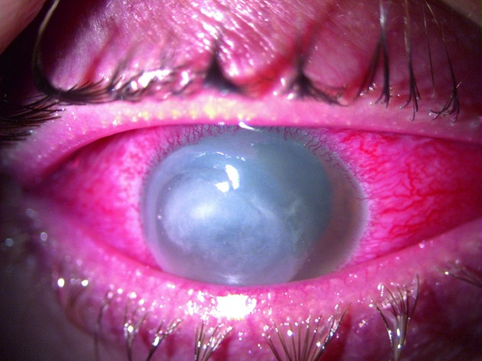
Interstitial keratitis manifests with varying degrees of corneal opacification and neovascularization. The neovascularization appears as the faint, pink areas in the peripheral cornea from the 11 o'clock to the 1 o'clock position.
© Springer Science+Business Media
Photophobia, pain, lacrimation, and vision blurring are common. The lesion begins as patches of inflammation in the mid-stroma that cause opacification. Typically with syphilis and occasionally with other causes, the entire cornea develops a ground-glass appearance, obscuring the iris. New blood vessels grow in from the limbus (neovascularization) and cause orange-red areas (salmon patches). Anterior uveitis and choroiditis are common in syphilitic interstitial keratitis. Inflammation and neovascularization usually begin to subside after 1 to 2 months regardless of treatment. Some corneal opacity usually remains, causing mild to moderate vision impairment.
Diagnosis of Interstitial Keratitis
Corneal opacification and other typical findings on slit-lamp examination
Serologic testing to determine etiology
The specific etiology must be determined. The stigmas of congenital syphilis, vestibuloauditory symptoms, history of an expanding rash, and tick exposure support specific etiologies. However, all patients should have serologic testing, including all of the following:
Fluorescent treponemal antibody absorption test or the microhemagglutination assay for Treponema pallidum are the usual syphilis screening tests
Lyme titer
Epstein-Barr virus panel
New tests such as Treponema pallidum particle agglutination assay, Treponema pallidum enzyme immunoassay, chemiluminescence immunoassay, and nucleic acid amplification tests (NAAT) may also be useful in diagnosing ocular syphilis.
Patients with negative serologic test results may have Cogan syndrome, an idiopathic syndrome consisting of interstitial keratitis and vestibular and auditory deficits. To prevent permanent vestibuloauditory damage, symptoms of hearing loss, tinnitus, or vertigo require urgent referral to an otolaryngologist.
Treatment of Interstitial Keratitis
Sometimes topical corticosteroids
Keratitis may resolve with treatment of the underlying condition. Additional topical treatment with a corticosteroid, such as prednisolone, is often advisable. An ophthalmologist should treat these patients.Keratitis may resolve with treatment of the underlying condition. Additional topical treatment with a corticosteroid, such as prednisolone, is often advisable. An ophthalmologist should treat these patients.
Key Points
Interstitial keratitis, which is rare in the United States, involves chronic inflammation of the middle corneal layers.
Findings include pain, tearing, decreased visual acuity, and often orange-red discoloration of the cornea and anterior uveitis.
Test patients for syphilis, Lyme disease, and Epstein-Barr virus infection.
Treatment is by an ophthalmologist; sometimes topical corticosteroids are prescribed.


