

Atypical nevi are benign melanocytic nevi with irregular and ill-defined borders; variegated colors, usually of brown and tan tones; and macular or papular components. Patients with atypical nevi have an increased risk of melanoma. Management is by close clinical monitoring and biopsy of highly atypical or changed lesions. Patients should reduce sun exposure and conduct regular self-examinations for new nevi or changes in existing ones.
Atypical nevi are nevi with a slightly different clinical and histologic appearance (disordered architecture and atypia of melanocytes). Some patients have only one or a few atypical nevi; others have many. Although most melanomas arise de novo, some develop from atypical nevi (1). Risk factors for melanoma include an increased number of atypical nevi and increased exposure to ultraviolet radiation and/or sunlight.

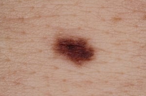
This image shows an atypical nevus, with the characteristic irregular border and variable coloration.
This image shows an atypical nevus, with the characteristic irregular border and variable coloration.
Image courtesy of Marie Schreiner, PA-C.

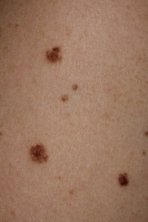
This image shows atypical nevi, with characteristic irregular borders and variable coloration.
This image shows atypical nevi, with characteristic irregular borders and variable coloration.
Image courtesy of Marie Schreiner, PA-C.
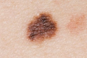
This photo shows a rounded mole with irregular borders.
This photo shows a rounded mole with irregular borders.
DR P. MARAZZI/SCIENCE PHOTO LIBRARY
This image shows an atypical nevus, with the characteristic irregular border and variable coloration.
This image shows an atypical nevus, with the characteristic irregular border and variable coloration.
Image courtesy of Marie Schreiner, PA-C.
This image shows atypical nevi, with characteristic irregular borders and variable coloration.
This image shows atypical nevi, with characteristic irregular borders and variable coloration.
Image courtesy of Marie Schreiner, PA-C.
This photo shows a rounded mole with irregular borders.
This photo shows a rounded mole with irregular borders.
DR P. MARAZZI/SCIENCE PHOTO LIBRARY
The propensity to develop atypical nevi may be inherited (autosomal dominant) or sporadic without apparent familial association. Familial atypical multiple mole–melanoma syndrome refers to the presence of multiple atypical nevi and melanoma in ≥ 2 first-degree relatives. Approximately 10% of patients with melanoma report a family history of melanoma, with CDKN2A mutations being the most common underlying mutation; however, incomplete penetrance that varies by geographic location is known to occur (2). These patients are at markedly increased risk of melanoma.
General references
1. Friedman RJ, Farber MJ, Warycha MA, et al: The "dysplastic" nevus. Clin Dermatol 27(1):103-115, 2009. doi: 10.1016/j.clindermatol.2008.09.008
2. Soura E, Eliades PJ, Shannon K, et al: Hereditary melanoma: Update on syndromes and management: Genetics of familial atypical multiple mole melanoma syndrome. J Am Acad Dermatol. 74(3):395-407, 2016; quiz 408-10. doi: 10.1016/j.jaad.2015.08.038
Symptoms and Signs of Atypical Nevi
Atypical nevi are often larger than other nevi (> 6 mm diameter) and primarily round (unlike many melanomas) but with indistinct borders and mild asymmetry. In contrast, melanomas have greater irregularity of color and may have areas that are red, blue, whitish, or depigmented with a scarred appearance.
Diagnosis of Atypical Nevi
Dermatoscopic evaluation
Biopsy
Regular physical examinations
Atypical nevi must be clinically differentiated from melanoma. Features that suggest melanoma, known as the ABCDEs of melanoma, are (1):
A: Asymmetry—asymmetric appearance
B: Borders—irregular borders (ie, not round or oval)
C: Color—color variation within the mole, unusual colors, or a color significantly different or darker than the patient's other nevi
D: Diameter—> 6 mm
E: Evolution—a new mole in a patient > 30 years of age or a changing mole
Although clinical findings can sometimes establish a diagnosis of atypical nevi (see table ), visual differentiation between atypical nevi and melanoma can be difficult. Therefore, biopsies of the most suspicious lesions should be performed to confirm the diagnosis and to determine the degree of atypia. Biopsies should ideally aim to include the complete depth and breadth of the lesion; an excisional biopsy is ideal.
Characteristics of Atypical vs Typical Nevi
Criteria | Typical Nevi | Atypical Nevi |
|---|---|---|
Age of onset | Childhood or adolescence | Continue to appear after adolescence |
Color | Flesh-colored, yellow-brown, or black | Tan to dark brown with a pink background; often resembling a fried egg, with a dark or light target commonly with a flatter rim than center Pigment often blurred at the edges or notched |
Diameter | 1–10 mm (usually < 6 mm) | 5–12 mm |
Shape | Symmetric with regular borders | Can be asymmetric or with irregular borders |
Location | Anywhere on the body | Most common on sun-exposed skin but may occur on covered areas (eg, buttocks, breasts, scalp) |
Number of lesions | ≤ 10 | One to several dozen |
Patients with multiple atypical nevi and a personal or family history of melanoma should be examined regularly (eg, yearly for those with a family history of melanoma, and more often for those with a personal history of melanoma). The familial risk of melanoma is also significantly increased by the presence of CDKN2A (p16) mutations, and to a lesser extent, BRCA2 mutations, particularly in the context of a family history of breast, ovarian, or pancreatic cancer (2, 3).
Some dermatologists observe pigment patterns of melanocytic lesions at higher resolutions using a hand-held instrument known as a dermatoscope. Dermoscopy allows a dermatologist to examine structures not typically visible to the naked eye. It can reveal certain high-risk characteristics suggestive of melanoma (eg, blue-white veil, irregular dots and globules, atypical pigment network, reverse network).
Diagnosis references
1. Rigel DS, Friedman RJ, Kopf AW, et al. ABCDE--an evolving concept in the early detection of melanoma. Arch Dermatol. 2005;141(8):1032-1034. doi:10.1001/archderm.141.8.1032
2. Potrony M, Puig-Butillé JA, Aguilera P, et al. Increased prevalence of lung, breast, and pancreatic cancers in addition to melanoma risk in families bearing the cyclin-dependent kinase inhibitor 2A mutation: implications for genetic counseling. J Am Acad Dermatol. 2014;71(5):888-895. doi:10.1016/j.jaad.2014.06.036
3. Gumaste PV, Penn LA, Cymerman RM, et al. Skin cancer risk in BRCA1/2 mutation carriers. Br J Dermatol. 2015;172(6):1498-1506. doi:10.1111/bjd.13626
Treatment of Atypical Nevi
Removal by excision or shave excision when desired
Complete excision of highly atypical lesions (sometimes also called "severely atypical") after histopathologic evaluation
Prophylactic removal of all atypical nevi is not effective in preventing melanoma and is not recommended. However, atypical nevi may warrant removal for any of the following conditions:
A patient has a high-risk history (eg, personal or family history of melanoma).
A patient cannot guarantee close follow-up.
The mole has high-risk dermatoscopic findings.
The mole is in a location that makes monitoring the lesion for changes difficult or impossible for the patient.
Highly or severely atypical nevi are moles that exhibit a high degree of abnormality and are difficult to distinguish from early melanoma under a dermatoscope. Multiple outcome studies have shown that the excision of mildly or moderately atypical nevi rarely results in a change in diagnosis, but the excision of severely atypical nevi (based on histopathologic tissue diagnosis) may detect melanoma in a small but clinically significant proportion of cases (1).
Treatment reference
1. Kim CC, Swetter SM, Curiel-Lewandrowski C, et al. Addressing the knowledge gap in clinical recommendations for management and complete excision of clinically atypical nevi/dysplastic nevi: Pigmented Lesion Subcommittee consensus statement. JAMA Dermatol. 2015;151(2):212-218. doi:10.1001/jamadermatol.2014.2694
Prevention of Atypical Nevi
Sun protection (ie, sun-protective clothing, sunscreen, sun avoidance during peak times, seeking shade)
Regular self-examination
Full-body photography
Sometimes surveillance of family members
Patients with atypical nevi should avoid excessive sun exposure and use sun protection. Patients who are vigilant about sun protection should be counseled to take sufficient supplemental vitamin D. Also, they should be taught self-examination to detect changes in existing nevi and to recognize features of melanomas. Full-body photography may help detect new nevi and monitor existing nevi for changes. Regular follow-up examinations are recommended.
If patients have a personal history of melanoma (whether developing from atypical nevi or de novo) or other skin cancers, first-degree relatives should also be referred for evaluation. Patients who are from melanoma-prone families (ie, ≥ 2 first-degree relatives with cutaneous melanomas) have a high lifetime risk of developing melanomas (1). To assess individual risk and plan appropriate follow-up, a comprehensive skin examination encompassing the entire skin surface (including the scalp and genitals) is recommended at least once for patients with high familial risk.
Prevention reference
1. Fallah M, Pukkala E, Sundquist K, et al. Familial melanoma by histology and age: joint data from five Nordic countries. Eur J Cancer. 2014;50(6):1176-1183. doi:10.1016/j.ejca.2013.12.023
Key Points
The risk of melanoma is higher if patients have increased numbers of atypical nevi, increased sun exposure, or familial atypical multiple mole–melanoma syndrome.
Because clinical differentiation from melanoma can be difficult, biopsy the most suspect atypical nevi.
Closely follow patients with atypical nevi, particularly those at higher risk of melanoma, and perform full-body photography.
Recommend sun protection (with supplemental vitamin D) and regular self-examination for high-risk patients.
Perform full-body examinations of all first-degree relatives of patients who have melanoma.


