

Detachment of the retina (the transparent, light-sensitive structure at the back of the eye) is separation of the retina from the underlying layer to which it is attached.
People notice a sudden increase in floaters, a sudden onset of flashing lights, a curtain or veil across vision, or sudden loss of vision.
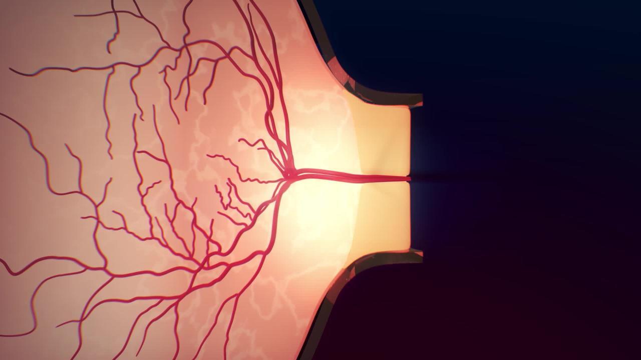
Doctors make the diagnosis by looking in the eye with an ophthalmoscope.
Most retinal detachments can be repaired, resulting in some restoration of vision if done soon after the detachment occurs.


A retinal detachment may begin in a small area, usually as the result of a retinal break (tear or, less commonly, a hole). If the small area is not soon reattached, the entire retina can detach. Retinal breaks that can lead to retinal detachment are more likely to occur in people who have or have had the following:
Severe nearsightedness (myopia)
An eye injury
Lattice retinal degeneration (a common disorder characterized by thinning and scar formation usually along the edge, or periphery, of the retina)
A family history of retinal detachment
When the retina detaches, it separates from part of its blood supply. Unless the retina is reattached, it may be permanently damaged by lack of blood. (See also Overview of Retinal Disorders.)
Sometimes a retinal detachment is not caused by a break. Some detachments are caused by complications of diseases that damage the retina (such as diabetes, which can cause diabetic retinopathy). Fluid or blood from a damaged blood vessel may also collect between the retina and the underlying tissue, causing a detachment.
Symptoms of a Detached Retina
A retinal detachment is painless. People usually see an increase in floating objects (floaters—objects that appear to move through a person's field of vision) or many flashes of bright light that last less than a second (photopsias) and have blurred vision. Peripheral vision is typically lost first, and vision loss spreads as the detachment progresses. The loss of vision causes grayness in the field of vision or resembles a curtain or veil falling across the line of sight.
People may have blood in the jellylike tissue (vitreous humor) near the back of the eye (vitreous hemorrhage). If the macula (the central area of the retina) becomes detached, vision rapidly deteriorates, and everything becomes blurred. Some retinal detachments do not cause symptoms at first.
Diagnosis of a Detached Retina
A doctor's examination of the eye
Sometimes ultrasonography
After applying eye drops to dilate the pupil, doctors examine the retina using an ophthalmoscope and can usually see a detachment. If the detachment is not visible, an ultrasound of the eye may help identify it.
Treatment of a Detached Retina
Surgical repair
For detachments caused by fluid leakage and without a retinal break, medications
Most retinal detachments can be repaired. The surgeon seals retinal breaks with laser surgery or freezing therapy (cryotherapy). For large retinal detachments, the surgeon may bring the retina and the wall of the eye together either by placing a silicone band around the eye (called a scleral buckle) or by removing the vitreous humor behind the lens and in front of the retina with surgery called a vitrectomy. A gas bubble is often used to hold the retina in place. For small detachments, laser surgery can prevent the retina from detaching more, or the retina can be reattached using cryotherapy plus a gas bubble (a procedure called pneumatic retinopexy).
Detachments that are caused by a disease that affects the retina (such as diabetes) can be treated with a vitrectomy.
Detachments that are caused by fluid leakage and that do not involve a retinal break may be treated with corticosteroids or medications that suppress the immune system (immunosuppressants, such as methotrexate and azathioprine) taken by mouth. Corticosteroids can also be given as an injected implant into the eye that slowly releases constant levels of a corticosteroid.Detachments that are caused by fluid leakage and that do not involve a retinal break may be treated with corticosteroids or medications that suppress the immune system (immunosuppressants, such as methotrexate and azathioprine) taken by mouth. Corticosteroids can also be given as an injected implant into the eye that slowly releases constant levels of a corticosteroid.
Prognosis for a Detached Retina
Surgery usually helps prevent additional vision loss. Vision often recovers except in the following circumstances:
The retina has been detached for several days or weeks.
Bleeding or scarring has occurred.
The macula has been detached or is damaged.
More Information
The following English-language resource may be useful. Please note that THE MANUAL is not responsible for the content of this resource.
National Eye Institute: A resource for learning about eye health (in English and Spanish) for adults and children, as well as for providing access to outreach campaigns. Simply type in the appropriate search term.


