

Acute external otitis is most commonly an infection of the ear canal skin that is typically caused by bacteria; Pseudomonas is the most common bacterial cause. Symptoms include pain, discharge, and hearing loss if the ear canal has swollen shut; manipulation of the auricle causes pain. Diagnosis is based on history and inspection. Treatment is with careful debridement of dead skin and wax, topical medications (including antibiotics, glucocorticoids, and acetic acid or a combination), and dry ear precautions. is the most common bacterial cause. Symptoms include pain, discharge, and hearing loss if the ear canal has swollen shut; manipulation of the auricle causes pain. Diagnosis is based on history and inspection. Treatment is with careful debridement of dead skin and wax, topical medications (including antibiotics, glucocorticoids, and acetic acid or a combination), and dry ear precautions.
Acute external otitis may manifest as a localized furuncle or as a diffuse infection of the entire canal (acute diffuse external otitis). The latter is often called swimmer’s ear; the combination of water in the ear canal and use of cotton swabs is the major risk factor. Malignant external otitis is a severe (usually due to Pseudomonas) osteomyelitis of the temporal bone, usually affecting older adults, patients with diabetes, and immunocompromised patients.
Acute external otitis is a common condition with an estimated lifetime prevalence of 10% (1).
General reference
1. Jackson EA, Geer K. Acute Otitis Externa: Rapid Evidence Review. Am Fam Physician. 2023;107(2):145-151.
Etiology of Acute External Otitis
Acute diffuse external otitis is usually caused by bacteria, such as Pseudomonas aeruginosa, Proteus vulgaris, Staphylococcus aureus, or Escherichia coli. Furuncles are usually caused by S. aureus and by methicillin-resistant S. aureus (MRSA). Fungal external otitis (otomycosis), typically caused by Aspergillus niger or Candida albicans, is less common.
Predisposing conditions include:
Inadvertent injury to the canal caused by cleaning with cotton swabs or other objects (eg, keys, hair pins)
Swimming
Allergies
Decreased canal acidity (possibly due to the repeated exposure to water)
Irritants (eg, hair spray, hair dye)
Use of earplugs or hearing aids (particularly if these devices are not adequately cleaned or do not fit correctly)
Disruptions in the skin-cerumen barrier (eg, even minor abrasions) contribute to the development of infection. Cerumen provides an acidic, hydrophobic barrier that naturally prevents the accumulation of water and, due to its low pH, prevents microbial growth. Attempts to clean the ear canal with cotton swabs can cause microabrasions of the delicate skin of the ear canal (these microabrasions act as portals of entry for bacteria) and may push debris and cerumen deeper into the canal. These accumulated substances tend to trap water, resulting in skin maceration that sets the stage for bacterial infection.
Symptoms and Signs of Acute External Otitis
Patients with acute external otitis have pain and drainage. Compared to chronic external otitis, acute external otitis is characterized by the rapid onset of symptoms (usually within 48 hours) (1). If the canal becomes swollen or filled with purulent debris, patients sometimes have a foul-smelling discharge and hearing loss.
The hallmark clinical sign of acute external otitis is exquisite tenderness that accompanies traction of the pinna or pressure over the tragus. Otoscopic examination in such patients is painful and difficult to conduct. The ear canal appears erythematous, swollen, and littered with moist, purulent debris and desquamated epithelium. In some cases, regional lymphadenopathy, cellulitis of the pinna, or perichondritis of the auricle may additionally be present.
Mild and moderate external otitis is characterized by ear pain, drainage, and hearing loss (nonsystemic symptoms isolated to the affected ear).
Severe external otitis is characterized by local symptoms including ear pain, drainage, hearing loss, and more systemic symptoms such as fever and fatigue; signs include possible regional lymphadenopathy, erythema and edema of the auricle.
Malignant external otitis (also called necrotizing otitis externa or skull base osteomyelitis) is characterized by high fever and the presence of granulation or necrotic tissue in the external ear canal, usually in a diabetic or immunocompromised patient (1). Extension to involve adjacent cranial nerves with subsequent neuropathy may occur. For more information, see Malignant External Otitis.

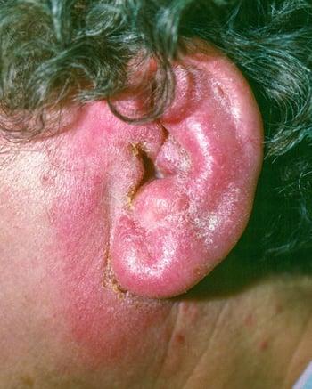
This image shows swelling, erythema, and dried discharge resulting from otitis externa accompanied by perichondritis of the auricle.
Otomycosis is more pruritic than painful, and patients also report aural fullness. Otomycosis caused by A. niger usually manifests with grayish black or yellow dots (fungal conidiophores) surrounded by a cottonlike material (fungal hyphae). In patients with infection caused by C. albicans, fungi are not visible, but usually, there is a thickened, creamy white cheesy exudate, which can be accompanied by spores that have a velvety appearance.

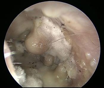
This image shows otomycosis of the external ear canal, as indicated by fungal hyphae and conidiophores of Aspergillus niger.
Furuncles cause severe pain and may drain sanguineous, purulent material. They appear as a focal, erythematous swelling (pimple).

In patients with otitis externa, the ear canal is erythematous and swollen and may be littered with purulent debris. As shown in this image, a furuncle (arrow) can develop in the infected canal.
Symptoms and signs reference
1. Jackson EA, Geer K. Acute Otitis Externa: Rapid Evidence Review. Am Fam Physician. 2023;107(2):145-151.
Diagnosis of Acute External Otitis
History and physical evaluation (including otoscopy)
Sometimes, additional testing (ie, imaging, culture)
The diagnosis of external otitis is typically based on inspection and eliciting a history of predisposing conditions. A clinical diagnosis is suspected in patients who present with all of the following (1):
Rapid onset of symptoms (generally within 48 hours) that have been present for less than 3 weeks
Inflammatory symptoms including severe otalgia, ear discharge, ear fullness (with or without hearing loss or jaw pain)
Inflammatory signs including tenderness of the tragus, pinna, or both, and/or diffuse ear canal edema or erythema (with or without otorrhea, regional lymphadenitis, or cellulitis of the pinna), and purulent drainage
Tympanometry (testing the mobility and function of the tympanic membrane) may be required for distinguishing acute otitis media from acute otitis externa, because systemic antimicrobial therapy may be required for the former (1). When discharge is copious, external otitis can be difficult to differentiate from an acute, purulent otitis media with tympanic membrane perforation. Pain elicited by pulling on the pinna suggests the presence of an external otitis.
Routine cultures are generally not necessary for uncomplicated acute external otitis as the diagnosis is frequently made clinically. Cultures can be considered for patients with persistent, recurrent, or severe infections, or in patients with immunocompromise (1). Fungal infections can be diagnosed by either appearance or culture.
In some cases, imaging (eg, CT or MRI) may be required to identify possible extension of infection into the skull base or bony involvement (malignant otitis externa).
Diagnosis reference
1. Rosenfeld RM, Schwartz SR, Cannon CR, et al. Clinical practice guideline: acute otitis externa. Otolaryngol Head Neck Surg. 2014;150(1 Suppl):S1-S24. doi:10.1177/0194599813517083
Treatment of Acute External Otitis
Debridement
Topical acetic acid and glucocorticoidsTopical acetic acid and glucocorticoids
Often topical antibiotics
The treatment of mild or moderate acute otitis externa consists of topical agents (primarily topical antimicrobials) for 7 to 10 days with analgesics for pain management (1, 2). No single topical preparation is clinically superior for initial treatment (3); therefore, selection is based on logistical, clinical, and patient factors (cost, tympanic membrane status, adherence, and adverse effect profiles). Systemic antibiotics are reserved for severe infection, spread beyond the canal, periauricular cellulitis, or in high-risk patients. Adjunctive approaches include thorough canal cleansing (debridement), avoiding water accumulation and further ear canal trauma (cessation of cotton-tipped applicators).
In mild or moderate acute external otitis, topical antibiotics and glucocorticoids are effective (eg, ciprofloxacin and fluocinolone) (topical antibiotics and glucocorticoids are effective (eg, ciprofloxacin and fluocinolone) (1, 4). First, the infected debris should be gently and thoroughly removed from the canal with suction or dry cotton swabs under adequate lighting and visualization. Water irrigation of the canal is absolutely contraindicated.
Mild external otitis can be treated by altering the ear canal’s pH with 2% acetic acid (or white vinegar) and by relieving inflammation with topical hydrocortisone; these are given as 5 drops 3 times a day for 7 days. Mild external otitis can be treated by altering the ear canal’s pH with 2% acetic acid (or white vinegar) and by relieving inflammation with topical hydrocortisone; these are given as 5 drops 3 times a day for 7 days.
Moderate external otitis requires the addition of an antibacterial solution or suspension, such as ciprofloxacin or ofloxacin. Moderate external otitis requires the addition of an antibacterial solution or suspension, such as ciprofloxacin or ofloxacin.Neomycin/polymyxin is no longer recommended because the neomycin component is highly sensitizing and causes an allergic reaction in up to 13% of patients (is no longer recommended because the neomycin component is highly sensitizing and causes an allergic reaction in up to 13% of patients (5). In addition, neomycin is ototoxic and risks injury to the inner ear and permanent hearing loss in the presence of a tympanic membrane perforation. When inflammation and swelling of the ear canal are relatively severe, an ear wick should be placed into the ear canal and wetted with Burow solution (5% aluminum acetate) or a topical antibiotic 4 times a day. The wick helps direct the drops deeper into the external canal when the canal is greatly swollen. The wick is left in place for 24 to 72 hours (or may fall out on its own); after this time, the swelling may have receded enough to allow drops to be instilled directly into the canal.). In addition, neomycin is ototoxic and risks injury to the inner ear and permanent hearing loss in the presence of a tympanic membrane perforation. When inflammation and swelling of the ear canal are relatively severe, an ear wick should be placed into the ear canal and wetted with Burow solution (5% aluminum acetate) or a topical antibiotic 4 times a day. The wick helps direct the drops deeper into the external canal when the canal is greatly swollen. The wick is left in place for 24 to 72 hours (or may fall out on its own); after this time, the swelling may have receded enough to allow drops to be instilled directly into the canal.
Severe external otitis or the presence of cellulitis extending beyond the ear canal may require systemic antibiotics (eg, ciprofloxacin 500 or 750 mg orally 2 times a day for 10 days). Quinolone antibiotics provide coverage against both extending beyond the ear canal may require systemic antibiotics (eg, ciprofloxacin 500 or 750 mg orally 2 times a day for 10 days). Quinolone antibiotics provide coverage against bothP. aeruginosa and S. aureus (1); however, their use is not recommended for children because tendon and cartilage damage is a risk (6). An analgesic, such as a nonsteroidal anti-inflammatory drug or even an oral opioid, may be necessary for the first 24 to 48 hours.
Fungal external otitis requires thorough cleaning of the ear canal and application of an antimycotic solution (eg, gentian violet, cresylate acetate, nystatin, clotrimazole, or even a combination of acetic acid and isopropyl alcohol). However, these solutions should not be used if the tympanic membrane is perforated because they can cause severe pain or damage the inner ear. Repeated cleanings and treatments may be needed to fully eradicate the infection. Reducing the relative humidity in the ear canal by avoiding hearing aid use and even blow drying the ear canal on a low setting to evaporate moisture and reduce humidity are also effective.requires thorough cleaning of the ear canal and application of an antimycotic solution (eg, gentian violet, cresylate acetate, nystatin, clotrimazole, or even a combination of acetic acid and isopropyl alcohol). However, these solutions should not be used if the tympanic membrane is perforated because they can cause severe pain or damage the inner ear. Repeated cleanings and treatments may be needed to fully eradicate the infection. Reducing the relative humidity in the ear canal by avoiding hearing aid use and even blow drying the ear canal on a low setting to evaporate moisture and reduce humidity are also effective.
A furuncle, if obviously pointing, should be incised and drained. However, if the patient is seen at an early stage, incision is of little value. Topical antibiotics are ineffective; oral antistaphylococcal antibiotics should be given. Analgesics may be necessary for pain relief. Dry heat can also lessen pain and hasten resolution.
In general, keeping the ear canal as dry as possible and reducing trauma (eg, from use of cotton tips) reduces the likelihood of infection. Dry ear precautions (eg, wearing a shower cap, avoiding swimming) are strongly advised for both external otitis and fungal external otitis. A blow dryer on a low setting can also be used to reduce existing humidity and moisture in the canal.
Pearls & Pitfalls
|
Treatment references
1. Rosenfeld RM, Schwartz SR, Cannon CR, et al. Clinical practice guideline: acute otitis externa. Otolaryngol Head Neck Surg. 2014;150(1 Suppl):S1-S24. doi:10.1177/0194599813517083
2. Jackson EA, Geer K. Acute Otitis Externa: Rapid Evidence Review. Am Fam Physician. 2023;107(2):145-151.
3. Kaushik V, Malik T, Saeed SR. Interventions for acute otitis externa. Cochrane Database Syst Rev. 2010;(1):CD004740. Published 2010 Jan 20. doi:10.1002/14651858.CD004740.pub2
4. Chu L, Acosta AM, Aazami H, et al. Efficacy and Safety of Ciprofloxacin Plus Fluocinolone Acetonide Among Patients With Acute Otitis Externa: A Randomized Clinical Trial. . Efficacy and Safety of Ciprofloxacin Plus Fluocinolone Acetonide Among Patients With Acute Otitis Externa: A Randomized Clinical Trial.JAMA Netw Open. 2022;5(7):e2221699. doi:10.1001/jamanetworkopen.2022.21699
5. Schapowal A. Contact Dermatitis to antibiotic ear drops is due to neomycin but not to ciprofloxacin. 5. Schapowal A. Contact Dermatitis to antibiotic ear drops is due to neomycin but not to ciprofloxacin.Allergy. 2001;56(suppl 68):14
6. Jackson MA, Schutze GE. The use of systemic and topical fluoroquinolones. Pediatrics. 2016;138 (5):e20162706. doi: 10.1542/peds.2016-2706
Prevention of Acute External Otitis
External otitis often can be prevented by applying a few drops of a 1:1 mixture of rubbing alcohol and white vinegar or acetic acid drops (as long as the eardrum is intact) immediately after swimming (External otitis often can be prevented by applying a few drops of a 1:1 mixture of rubbing alcohol and white vinegar or acetic acid drops (as long as the eardrum is intact) immediately after swimming (1). The alcohol helps remove (evaporate) water, and the vinegar alters the pH of the canal. Use of cotton swabs or other implements in the canal should be strongly discouraged.
Prevention reference
1. Jackson EA, Geer K. Acute Otitis Externa: Rapid Evidence Review. Am Fam Physician. 2023;107(2):145-151.
Key Points
Acute external otitis is usually bacterial (pseudomonal); fungal infections are less common and cause more itching and less pain.
Severe pain when the pinna is pulled suggests acute external otitis.
Under close and direct visualization, gently remove infected debris from the canal with suction or dry cotton swabs.
Do not irrigate the ear.
For mild cases, apply acetic acid and hydrocortisone drops.For mild cases, apply acetic acid and hydrocortisone drops.
For moderate or severe cases, debridement and topical antibiotics (use a wick if the canal is swollen) are critical; sometimes give systemic antibiotics.
More Information
The following English-language resource may be useful. Please note that The Manual is not responsible for the content of this resource.
Hajioff D, MacKeith S. Otitis externa. BMJ Clin Evid. 2015;0510. Published 2015 Jun 15.
Drug Information for the Topic


