

Oculocutaneous albinism is an inherited defect in melanin formation that causes diffuse hypopigmentation of the skin, hair, and eyes. Ocular albinism affects the eyes and usually not the skin. Ocular involvement causes photophobia, strabismus, nystagmus, and decreased vision. Diagnosis of oculocutaneous albinism is usually obvious from the skin examination, but ocular evaluation is necessary. No treatment for the skin involvement is available other than protection from sunlight.
(See also Overview of Pigmentation Disorders.)
Pathophysiology of Albinism

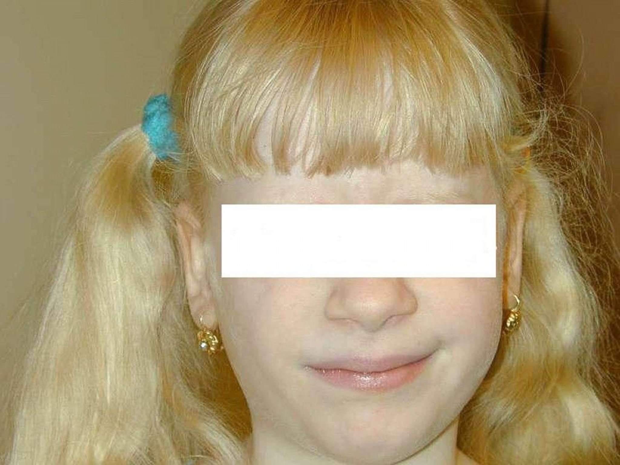
Photo courtesy of Noah S. Scheinfeld, MD, and the Dermatology Online Journal.
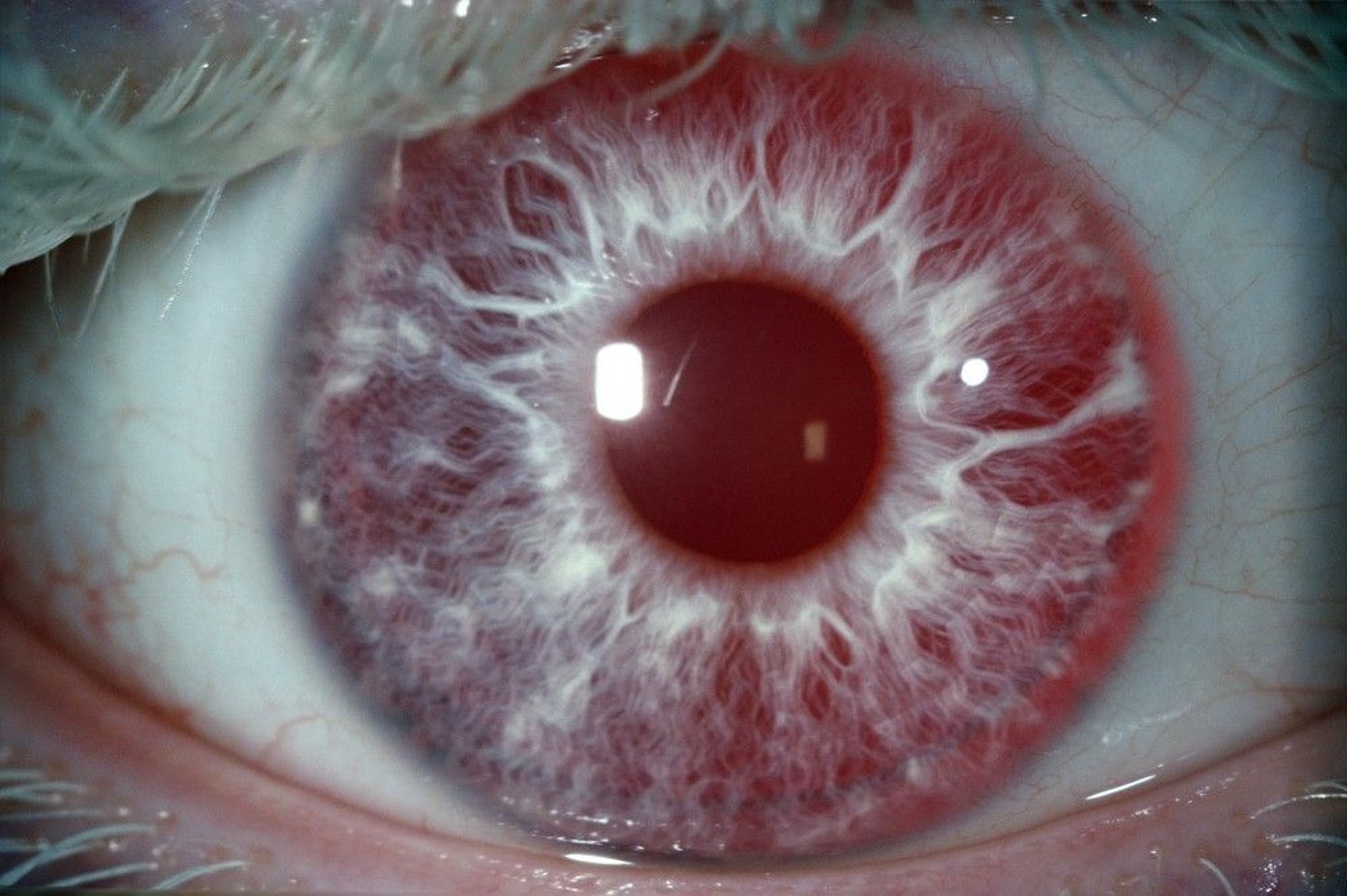
Oculocutaneous albinism (OCA) is a group of rare inherited disorders in which a normal number of melanocytes are present but melanin production is absent or greatly decreased. OCA occurs in people of all races throughout the world. Cutaneous and ocular pathologies (ocular involvement) are both present. Findings in ocular involvement include abnormal optic tract development manifested by foveal hypoplasia with decreased photoreceptors and misrouting of optic chiasmal fibers.

BSIP, KOKEL/SCIENCE PHOTO LIBRARY
Most cases of OCA are autosomal recessive; autosomal dominant inheritance is rare. There are 8 genetic forms of OCA, of which the first 4 are well characterized. See table .
Types and Phenotypes of Oculocutaneous Albinism (OCA)
Type (OMIM Number) | Defect or Mutation | Phenotypes | Comments |
|---|---|---|---|
Type I | Mutations in TYR gene result in absent (OCA1A) or reduced (OCA1B) tyrosinase activity. Tyrosinase catalyzes several steps in melanin synthesis. | In OCA1A, skin and hair are milky white, and eyes are blue-gray (decrease in visual acuity is the most severe in this form of OCA). In OCA1B, pigmentary dilution ranges from obvious to subtle. | OCA1A comprises 40% of all OCA. |
Type II (203200) | P (pink-eyed) gene The function of the P protein is not yet known but may involve regulation of organelle pH and accumulation of vacuolar glutathione. | OCA type II has phenotypes with pigmentary dilution that ranges from minimal to moderate. Pigmented nevi and lentigines may develop if skin is exposed to the sun; some lentigines become large and dark. Eye color varies greatly. | Tyrosinase activity is present. Type II comprises 50% of all OCA and is the most common form of OCA in Africa. |
Type III (203290) | Tyrosinase-related protein 1 (TYRP1) gene The product of this gene is important in the synthesis of eumelanin, the most common of the 3 types of melanin that give skin, hair, and eyes their color. | In OCA type III, skin is brown, hair is rufous (reddish), and eye color can be blue or brown. | Type III occurs only in people with otherwise dark skin (Fitzpatrick skin types III to V—see table Fitzpatrick Skin Type Classification). |
Type IV (606574) | SLC45A2 gene This gene codes a membrane transporter protein involved in tyrosinase processing and trafficking of proteins to melanosomes. | In OCA type IV, the phenotype is similar to that for type II. | Type IV is an extremely rare form. It is the most common form of OCA in Japan. |
Type V (615312) | Linked to chromosome 4q24 This chromosome is located in a region that may code for lysosomal proteins. | In OCA type V, skin is white, and hair is golden-colored. | — |
Type VI (113750) | Linked to mutations in gene SLC45A5 This gene encodes a membrane transporter protein. | In OCA type VI, skin can be white, and hair can be light at birth and may darken with age. | — |
Type VII (615179) | Mutations in gene C10ORF11 This gene encodes a leucine-rich protein that may play a role in melanocyte differentiation. | In OCA type VII, skin pigment is decreased, and hair can range from white to brown. | — |
Type VIII (619165) | Linked to a mutation in gene dopachrome tautomerase (DCT) on chromosome 13q32 This gene encodes an enzyme that plays a role in modification of skin pigment color. | In OCA type VIII, skin and hair are mildly hypopigmented. | — |
Nettleship-Falls (ocular albinism 1 [OA1]) and Forsius-Eriksson (OA2) are extremely rare compared to OCA. They are inherited in an X-linked dominant fashion. Usually findings are confined to the eyes, but skin may be hypopigmented. Patients with OA1 may have late-onset sensorineural deafness.
In another group of inherited diseases, a clinical phenotype of OCA occurs in conjunction with bleeding disorders. In Hermansky-Pudlak syndrome, OCA-like findings occur with platelet abnormalities and a ceroid-lipofuscin lysosomal storage disease (which can lead to pulmonary fibrosis and granulomatous colitis). This syndrome is rare except in people with family origin in Puerto Rico, where its incidence is approximately 1 in 1800 (1). In Chédiak-Higashi syndrome, OCA-like cutaneous and ocular findings occur, hair is silvery gray, and a decrease in platelet-dense granules results in a bleeding diathesis. Patients with Chédiak-Higashi syndrome have severe immunodeficiency due to abnormal lymphocyte lytic granules and progressive neurologic degeneration.
General reference
1. Witkop CJ, Almadovar C, Piñeiro B, et al : Hermansky-Pudlak syndrome (HPS). An epidemiologic study. Ophthalmic Paediatr Genet 11(3):245-250. doi: 10.3109/13816819009020986
Symptoms and Signs of Albinism
The different genetic forms of oculocutaneous albinism have a variety of phenotypes. See table .
Patients with ocular involvement may have decreased retinal pigmentation, leading to extreme sensitivity to light (photophobia) and light avoidance. In addition, nystagmus, strabismus, reduced visual acuity, and loss of binocular stereopsis likely result from defective routing of the optic fibers.
Diagnosis of Albinism
Clinical evaluation
Diagnosis of all types of OCA and OA is based on examination of the skin and eyes. Early ocular examination may detect iris translucency, reduced retinal pigmentation, foveal hypoplasia, reduced visual acuity, strabismus, and nystagmus.
Because it may not always be possible to distinguish the subtypes of OCA, genetic testing may be helpful.
Treatment of Albinism
Strict sun protection
Sometimes surgical intervention for strabismus
There is no cure for albinism.
Patients are at high risk of sunburn and skin cancers (especially squamous cell carcinoma) and should avoid direct sunlight, use sunglasses with ultraviolet (UV) filtration, wear sun-protective clothing with a UV protection factor of 50 or higher, and use sunscreen with a broad-spectrum sun protection factor (SPF) of 50 or higher that protects against UVA and UVB wavelengths (see sun exposure prevention).
Patients with OCA should have regular skin examinations. Of note, melanoma that develops in patients with OCA is frequently amelanotic; common sites include the back and legs.
Key Points
Oculocutaneous albinism is a group of rare, usually autosomal recessive disorders, resulting in hypopigmentation of the skin, hair, and eyes.
Ocular involvement causes photosensitivity and often nystagmus, strabismus, reduced visual acuity, and loss of binocular stereopsis.
Examine the eyes and skin to make the diagnosis.
Instruct patients on how to strictly protect the skin and eyes from sun exposure.


