

Nummular dermatitis is inflammation of the skin characterized by coin-shaped or discoid eczematous lesions. Diagnosis is clinical. Treatment may include topical corticosteroids and phototherapy.
(See also Definition of Dermatitis.)
Many patients with nummular dermatitis are atopic. In these cases, nummular dermatitis is simply a localized manifestation of atopic dermatitis (nummular atopic dermatitis). Patients with atopic dermatitis can have nummular plaques side-by-side with other, more common manifestations of atopic dermatitis. However, some patients with nummular dermatitis do not have atopy. In these patients, the etiology is unclear.
Bacterial colonization or allergic contact reactions (1), either at the site of the lesions or elsewhere (autoeczematization or id reaction, dermatitis at sites remote from the site of the initial inflammatory problem or infection) are possible causes.
Nummular dermatitis is most common among middle-aged and older patients.
General reference
1. Bonamonte D, Foti C, Vestita M, et al. Nummular eczema and contact allergy: A retrospective study. Dermatitis. 23(4):153–157, 2012. doi: 10.1097/DER.0b013e318260d5a0
Symptoms and Signs of Nummular Dermatitis
Plaques and patches of nummular dermatitis are erythematous and scaly, typically intensely pruritic, and are coin-shaped and well-demarcated but not sharply. They can number from 1 to approximately 50 and tend to be from 2 to 10 cm in diameter. They are often more prominent on the extensor aspects of the extremities and on the buttocks but also appear on the trunk.

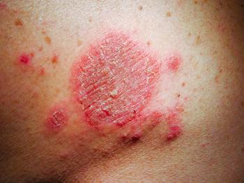
This photo shows discoid, erythematous, and scaly lesions consistent with nummular dermatitis in light skin.


This photo shows a thin, well- but not sharply demarcated lichenified and hyperpigmented plaque. Erythema is not discernible, likely because of the dark skin color.
Photo courtesy of Karen McKoy, MD.
Diagnosis of Nummular Dermatitis
Clinical evaluation
Diagnosis of nummular dermatitis is clinical and based on the characteristic appearance and distribution of the skin lesions. Patients should be evaluated for atopy.
In nonatopic patients, allergic contact dermatitis should be considered, and patch testing may be helpful.
Differential diagnoses include:
Treatment of Nummular Dermatitis
Supportive care, including counseling
Antipruritics
Corticosteroids (most often topical)
Phototherapy
Treatment of nummular dermatitis is similar to that of atopic dermatitis and includes counseling, antipruritics, corticosteroids, and sometimes phototherapy (particularly narrowband ultraviolet B).
Dupilumab, topical calcineurin inhibitors (tacrolimus and pimecrolimus), and/or crisaborole should be considered for nummular atopic dermatitis. Dupilumab, topical calcineurin inhibitors (tacrolimus and pimecrolimus), and/or crisaborole should be considered for nummular atopic dermatitis.
Rarely, systemic immunosuppressants are required.
Key Points
Nummular dermatitis is often a manifestation of atopic dermatitis; in nonatopic patients, the etiology of nummular dermatitis is unknown.
Patients present with single or multiple pruritic coin-shaped, well-demarcated, erythematous scaly patches or plaques.
Diagnosis is clinical; skin infections and psoriasis must be excluded.
Treatment includes topical corticosteroids and phototherapy; systemic immunosuppressants are rarely needed.


