

Brain aneurysms are focal dilations in the cerebral arteries.
In the United States, brain aneurysms occur in 3 to 5% of people (1). Brain aneurysms can occur at any age but are most common among people aged 30 to 60 years and are more common among women than men.
Common contributing factors for aneurysms may include:
Hereditary connective tissue disorders (eg, Ehlers-Danlos syndrome, pseudoxanthoma elasticum, autosomal dominant polycystic kidney syndrome)
Family history of aneurysm (first-degree relative: parent, sibling, or child)
Cigarette smoking
Polysubstance abuse
Amyloid deposition
Arteriovenous malformations
Occasionally, septic emboli cause mycotic aneurysms.
Hypertension weakens the wall of the aneurysm by mechanical stress and causes vascular inflammation; both effects contribute to aneurysm rupture. Blood pressure control is important in preventing aneurysm rupture.
Brain aneurysms are most often < 2.5 cm in diameter and saccular (noncircumferential); sometimes they have one or more small, thin-walled, outpouchings (berry aneurysm).
Most brain aneurysms occur along the middle or anterior cerebral arteries or the communicating branches of the circle of Willis, particularly at arterial bifurcations. Mycotic aneurysms usually develop distal to the first bifurcation of the arterial branches of the circle of Willis.
General reference
1. Vlak MH, Algra A, Brandenburg R, Rinkel GJ. Prevalence of unruptured intracranial aneurysms, with emphasis on sex, age, comorbidity, country, and time period: a systematic review and meta-analysis. Lancet Neurol. 2011;10(7):626-636. doi:10.1016/S1474-4422(11)70109-0
Symptoms and Signs of Brain Aneurysms
Many aneurysms are asymptomatic, but a few, usually large or growing aneurysms, cause symptoms by compressing adjacent structures. Ocular palsies, diplopia, squint, or orbital pain may indicate pressure on the 3rd, 4th, 5th, or 6th cranial nerves. Visual loss and a bitemporal field defect may indicate pressure on the optic chiasm.
Brain aneurysms may bleed into the subarachnoid space, causing subarachnoid hemorrhage or at times intracerebral hemorrhage. Before rupture, aneurysms occasionally cause sentinel (warning) headaches due to painful expansion of the aneurysm or to blood leaking into the subarachnoid space. Actual rupture causes a sudden severe headache called a thunderclap headache.
A ruptured aneurysm may also cause nausea, vomiting, a stiff neck, photosensitivity, loss of consciousness, and/or seizures.
Diagnosis of Brain Aneurysms
Neuroimaging
Neuroimaging may detect aneurysms incidentally.
Diagnosis of aneurysms requires angiography, CT angiography, or magnetic resonance angiography. Catheter-based digital subtraction angiography (DSA) is the gold standard for diagnosing aneurysms. When initial CT angiography or DSA is negative for an aneurysm in patients with subarachnoid hemorrhage around the circle of Willis, delayed DSA performed 7 days after the initial aneurysmal bleeding is valuable.
If a mycotic aneurysm is suspected, bacterial and fungal blood cultures should be performed.

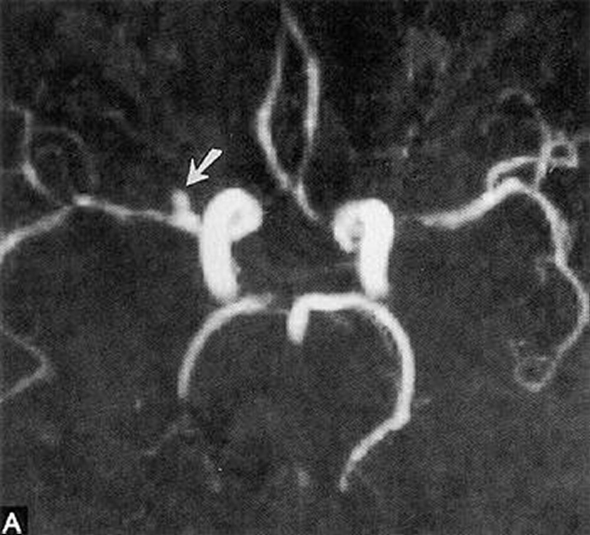
By permission of the publisher. From Ritter A, Hayman L, Charletta D. In Atlas of Cerebrovascular Disease. Edited by PB Gorelick and MA Sloan. Philadelphia, Current Medicine, 1996.
Treatment of Brain Aneurysms
For smaller, asymptomatic aneurysm, serial imaging
For large or symptomatic aneurysms, endovascular therapy
Treatment of unruptured aneurysms depends on
Type, size, and location of the aneurysm
Risk of rupture
Patient age and health
Personal and family medical history
Risks of treatment
Risk of rupture versus that of perioperative complications should be discussed frankly with the patient.
Control of atherosclerotic risk factors, especially smoking cessation and use of antihypertensive medications as appropriate, is important.
If < 7 mm, asymptomatic aneurysms in the anterior circulation rarely rupture and do not warrant the risks of immediate treatment. They can be monitored with serial imaging.
If aneurysms are larger, are in the posterior circulation, or cause symptoms due to bleeding or compression of neural structures, endovascular therapy (eg, stents, coil embolization), if feasible, can be tried. Sometimes open surgery with placement of a microvascular clip is necessary.
Early repair is essential if patients have had aneurysmal bleeding because rebleeding is the most serious early complication with fatality rate up to 70%.
Treatment of mycotic aneurysms is aggressive antibiotic therapy directed at the specific pathogen. Usually, mycotic aneurysms must also be surgically repaired.
If the aneurysm has ruptured, digital subtraction angiography is used to locate it; then endovascular therapy or open surgery is performed to repair it.
Key Points
Before rupture, aneurysms occasionally cause sentinel (warning) headaches; actual rupture causes a sudden severe headache (thunderclap headache).
Diagnose using angiography, CT angiography, or magnetic resonance angiography.
If the aneurysm is asymptomatic and < 7 mm, monitor with serial imaging; if the aneurysm is symptomatic, larger, and located in the posterior circulation, treat using endovascular therapy or sometimes open surgery.
If the aneurysm has ruptured, use digital subtraction angiography to locate the aneurysm, then proceed with endovascular therapy or open surgery.


