

Erysipeloid is an infection caused by the gram-positive bacillus Erysipelothrix rhusiopathiae. The most common manifestation is an acute but slowly evolving localized cellulitis. Diagnosis is by culture of a biopsy specimen or occasionally polymerase chain reaction testing. Treatment is with antibiotics.
Erysipeloid is an infection caused by the bacterium Erysipelothrix rhusiopathiae, which is a thin, gram-positive, capsulated, nonsporulating, nonmotile, aerobic or facultatively anaerobic bacillus with worldwide distribution; they are primarily saprophytes (ie, living on dead and/or decaying matter).
E. rhusiopathiae may infect a variety of animals, including shellfish, fish, birds, and mammals (especially swine), and insects. In humans, infection is a zoonosis, chiefly occupational, and typically follows a penetrating wound in people who handle edible or nonedible animal matter (eg, infected carcasses, rendered products [grease, fertilizer], bones, shells). Most commonly, patients handle fish or shellfish or work in slaughterhouses handling raw meat where associated abrasions or puncture wounds serve as sites of entry in most cases of infection. Infection can also result from cat or dog bites.
Pathophysiology of Erysipeloid
E. rhusiopathiae is an encapsulated organism with virulence factors capable of resisting phagocytosis and supporting intracellular survival, particularly within macrophages (1). Key virulence factors include surface protective antigen A (SpaA), an adhesion protein that enhances attachment to host cells and has shown protective effects in preclinical vaccination models (2). Additional virulence factors include enzymes such as neuraminidase and hyaluronidase, as well as various cell wall-associated proteins. ). Additional virulence factors include enzymes such as neuraminidase and hyaluronidase, as well as various cell wall-associated proteins.
Virulence factors contribute to the failure of host immune surveillance and allow the organism to spread systemically once a cutaneous barrier has been breached, affecting multiple organ systems, including the cardiovascular system, central nervous system, kidneys, joints, and lungs. Extracutaneous infections are rare, however, and usually manifest as septic arthritis or infective endocarditis.
Pathophysiology references
1. Shimoji Y. Pathogenicity of Erysipelothrix rhusiopathiae: virulence factors and protective immunity. Microbes Infect. 2000;2(8):965-972. doi:10.1016/s1286-4579(00)00397-x
2. Cheun HI, Kawamoto K, Hiramatsu M, et al. Protective immunity of SpaA-antigen producing Lactococcus lactis against Erysipelothrix rhusiopathiae infection. J Appl Microbiol. 2004;96(6):1347-1353. doi:10.1111/j.1365-2672.2004.02283.x
Symptoms and Signs of Erysipeloid
Erysipeloid infections may cause localized or generalized cutaneous lesions. Some patients develop systemic manifestations, some of whom may also develop endocarditis.
Localized cutaneous infection: Within 1 week of injury, a slowly evolving characteristic raised, violaceous, indurated, localized cellulitic rash appears on the affected area (eg, hand), accompanied by itching and burning. Local swelling, although sharply demarcated, may inhibit use of the hand, the usual site of infection. The lesion’s border may slowly extend outward, causing discomfort and disability that may persist for 3 weeks. Localized erysipeloid is usually self-limited. Regional lymphadenopathy and lymphangitis can occur in about one-third of cases (1).
Generalized cutaneous infection: Erysipeloid rarely becomes a generalized cutaneous disease, which is characterized by violaceous skin lesions that expand as the lesion’s center clears, plus bullous lesions at the primary or distant sites. Adjoining lymphangitis may also occur. Systemic symptoms are generally absent.
Systemic infection with or without endocarditis: Bacteremia is rare and is more often a primary infection than dissemination from cutaneous lesions. It may result in septic arthritis or infective endocarditis, even in people without known valvular heart disease. Endocarditis tends to involve the aortic valve, and the mortality rate and percentage of patients needing cardiac valve replacement are unusually high (2, 3). Rarely, central nervous system, intra-abdominal, and bone infections occur.

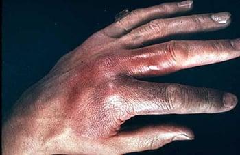
This image shows the characteristic violaceous, indurated rash of erysipeloid infection.
Symptoms and signs references
1. Stevens DL, Bisno AL, Chambers HF, et al. Practice guidelines for the diagnosis and management of skin and soft tissue infections: 2014 update by the Infectious Diseases Society of America. Clin Infect Dis. 2014;59(2):147-159. doi:10.1093/cid/ciu296
2. Balkhair A, Al Lawati H, Al Riyami M, Alameddine T, Al Amin M, Al Adawi B. Erysipelothrix rhusiopathiae endocarditis diagnosed by broad range 16s rRNA PCR gene sequencing. IDCases. 2019;18:e00584. doi:10.1016/j.idcr.2019.e00584
3. Rostamian M, Rahmati D, Akya A. Clinical manifestations, associated diseases, diagnosis, and treatment of human infections caused by Erysipelothrix rhusiopathiae: a systematic review. Germs. 2022;12(1):16-31. doi:10.18683/germs.2022.1303
Diagnosis of Erysipeloid
Culture
Polymerase chain reaction (PCR) amplification for rapid diagnosis
Culture of a full-thickness biopsy specimen is superior to needle aspiration of the advancing edge of a lesion because organisms are located only in deeper parts of the skin. Culture of exudate obtained by abrading a florid papule may be diagnostic. Isolation from synovial fluid or blood is necessary for the diagnosis of arthritis or endocarditis due to E. rhusiopathiae infection, however, blood cultures are rarely positive (1). E. rhusiopathiae may be misidentified as lactobacilli or enterococcus (2).
PCR-based amplification assays may aid in the rapid diagnosis of erysipeloid. Rapid diagnosis is particularly important if endocarditis is suspected because treatment of endocarditis due to E. rhusiopathiae is often different from the usual empiric treatment of gram-positive bacillary endocarditis (eg, E. rhusiopathiae is resistant to vancomycin, which is typically used).is resistant to vancomycin, which is typically used).
Diagnosis references
1. Stevens DL, Bisno AL, Chambers HF, et al. Practice guidelines for the diagnosis and management of skin and soft tissue infections: 2014 update by the Infectious Diseases Society of America. Clin Infect Dis. 2014;59(2):147-159. doi:10.1093/cid/ciu296
2. Dunbar SA, Clarridge JE 3rd. Potential errors in recognition of Erysipelothrix rhusiopathiae. J Clin Microbiol. 2000;38(3):1302-1304. doi:10.1128/JCM.38.3.1302-1304.2000
Treatment of Erysipeloid
Penicillin, imipenem, cephalosporins, fluoroquinolones or clindamycin
Infection is treated with antibiotics (1). The choice and dosage protocol of antibiotic depends on the location of the infection.
For localized cutaneous disease, usual treatment is one of the following, given for 7 to 10 days (1):
Penicillin V or ampicillin (500 mg orally every 6 hours) or amoxicillin (500 mg orally every 8 hours)Penicillin V or ampicillin (500 mg orally every 6 hours) or amoxicillin (500 mg orally every 8 hours)
Ciprofloxacin (250 mg orally every 12 hours)Ciprofloxacin (250 mg orally every 12 hours)
Clindamycin (300 mg orally every 8 hours)Clindamycin (300 mg orally every 8 hours)
Cephalosporins are also effective. Daptomycin and linezolid are active in vitro and may be considered if patients are very allergic to beta-lactams. Tetracyclines and macrolides may no longer be dependable.Cephalosporins are also effective. Daptomycin and linezolid are active in vitro and may be considered if patients are very allergic to beta-lactams. Tetracyclines and macrolides may no longer be dependable.
E. rhusiopathiae are resistant to sulfonamides, aminoglycosides, and vancomycin.
Severe diffuse cutaneous or systemic infection is best treated with one of the following:
Penicillin G (2 to 3 million units IV every 4 hours)Penicillin G (2 to 3 million units IV every 4 hours)
Ceftriaxone (2 g IV once a day)Ceftriaxone (2 g IV once a day)
A fluoroquinolone (eg, ciprofloxacin 400 mg IV every 12 hours, levofloxacin 500 mg IV once a day)A fluoroquinolone (eg, ciprofloxacin 400 mg IV every 12 hours, levofloxacin 500 mg IV once a day)
Endocarditis is treated with penicillin G for 4 to 6 weeks. Cephalosporins and fluoroquinolones are alternatives. Vancomycin is often used empirically for the treatment of gram-positive bacillary endocarditis; however, is treated with penicillin G for 4 to 6 weeks. Cephalosporins and fluoroquinolones are alternatives. Vancomycin is often used empirically for the treatment of gram-positive bacillary endocarditis; however,E. rhusiopathiae is resistant to vancomycin. Thus, rapid differentiation of E. rhusiopathiae from other gram-positive organisms is critical to initiate appropriate antibiotic therapy.
Arthritis is treated with the same antibiotics and doses as endocarditis (given for at least 1 week after defervescence or cessation of effusion), but repeated needle aspiration drainage of the infected joint is also necessary.
Treatment reference
1. Stevens DL, Bisno AL, Chambers HF, et al. Practice guidelines for the diagnosis and management of skin and soft tissue infections: 2014 update by the Infectious Diseases Society of America. Clin Infect Dis. 2014;59(2):147-159. doi:10.1093/cid/ciu296
Key Points
Erysipeloid typically results from a penetrating wound in people who handle edible or nonedible animal matter (eg, in a slaughterhouse) or who work with fish or shellfish.
Within 1 week after the injury, a raised, violaceous, nonvesiculated, indurated, maculopapular rash appears, accompanied by itching and burning; about one-third of patients have lymphangitis or regional lymphadenopathy.
Bacteremia is rare but may result in septic arthritis or infective endocarditis.
Diagnosis is based on culturing a full-thickness biopsy of a skin specimen or an exudate obtained by abrading a florid papule.
If endocarditis due to E. rhusiopathiae is suspected, rapid identification of the pathogen is critical because treatment is often different from the usual empiric treatment of gram-positive bacillary endocarditis; E. rhusiopathiae is resistant to vancomycin, which is typically used to treat gram-positive bacillary endocarditis.
Treat with antibiotics (eg, penicillin, ciprofloxacin) based on the extent and location of infection.


