


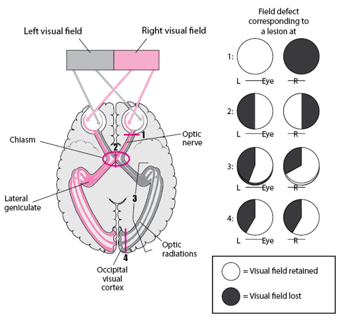
The optic pathway includes the retina, optic nerve, optic chiasm, optic radiations, and occipital cortex (see figure ). Damage along the optic pathway causes a variety of visual field defects. The type of field defect can help localize the lesion (see table ).
Higher Visual Pathways—Lesion Sites and Corresponding Visual Field Defects
With retrochiasmal lesions, visual field defects are on the same side (homonymous) and tend to become more symmetric (congruous) with more posterior lesions, as shown with the occipital lesion in #4. |

Types of Visual Field Defects
Type | Description | Causes |
|---|---|---|
Altitudinal field defect | Loss of all or part of the superior or inferior half of the visual field; does not cross the horizontal median | More common: Ischemic optic neuropathy (usually nonarteritic), hemibranch retinal artery occlusion, retinal detachment Less common: Glaucoma, optic neuritis, optic nerve coloboma |
Arcuate scotoma | A small, bow-shaped (arcuate) visual field defect that follows the arcuate pattern of the retinal nerve fibers; does not cross the horizontal median | Damage to ganglion cells that feed into a particular part of the optic nerve head More common: Glaucoma Less common: Ischemic optic neuropathy (usually nonarteritic), papilledema, optic disc drusen, high myopia |
Binasal field defect (uncommon) | Loss of all or part of the medial half of both visual fields; does not cross the vertical median | More common: Glaucoma, bitemporal retinal disease (eg, retinitis pigmentosa) Rare: Tumor or aneurysm compressing both optic nerves |
Bitemporal hemianopia | Loss of all or part of the lateral half of both visual fields; does not cross the vertical median | More common: Chiasmal lesion (eg, pituitary adenoma, meningioma, craniopharyngioma, aneurysm, glioma) Less common: Tilted optic discs Rare: Binasal retinal disease (eg, retinitis pigmentosa or acute zonal occult outer retinopathy [AZOOR]) |
Blind-spot enlargement | Enlargement of the normal blind spot at the optic nerve head | Papilledema, optic nerve drusen, optic nerve coloboma, myelinated nerve fibers at the optic disc, drugs and medications, myopic disc with a crescent, AZOOR, multiple evanescent white dot syndrome (MEWDS), acute idiopathic blind spot enlargement (AIBSE) syndrome |
Central scotoma | A loss of visual function in the middle of the visual field | Macular disease, optic neuropathy (eg, optic neuritis, compressive optic neuropathy, Leber hereditary optic neuropathy, vitamin deficiency, or toxic-metabolic disorders) Rare: Occipital cortex lesion |
Constriction of the peripheral fields, leaving only a small residual central field | Loss of the outer part of the entire visual field in one or both eyes | Glaucoma, retinitis pigmentosa or other peripheral retinal disorder, chronic papilledema, panretinal photocoagulation, central retinal artery occlusion with cilioretinal artery sparing, bilateral occipital lobe infarction with macular sparing, cancer-associated retinopathy, autoimmune retinopathy, functional vision loss Rare: Medications (eg, hydroxychloroquine) |
Homonymous hemianopia | Loss of part or all of the left half or right half of both visual fields; does not cross the vertical median | Lesion anywhere posterior to the optic chiasm: optic tract or lateral geniculate body lesion; lesion in temporal, parietal, or occipital lobe (more commonly, stroke or tumor; less commonly, aneurysm or trauma); migraine* (which may cause transient homonymous hemianopia) |
* Migraine can cause various transient visual field defects, although it most commonly causes a transient homonymous hemianopia. | ||
Adapted from Gervasio KA, Peck TJ, Fathy CA, et al.: The Wills Eye Manual: Office and Emergency Room Diagnosis and Treatment of Eye Disease, ed. 8. Lippincott, Williams &Wilkins, a Wolters Kluwer business; 2022. | ||


