


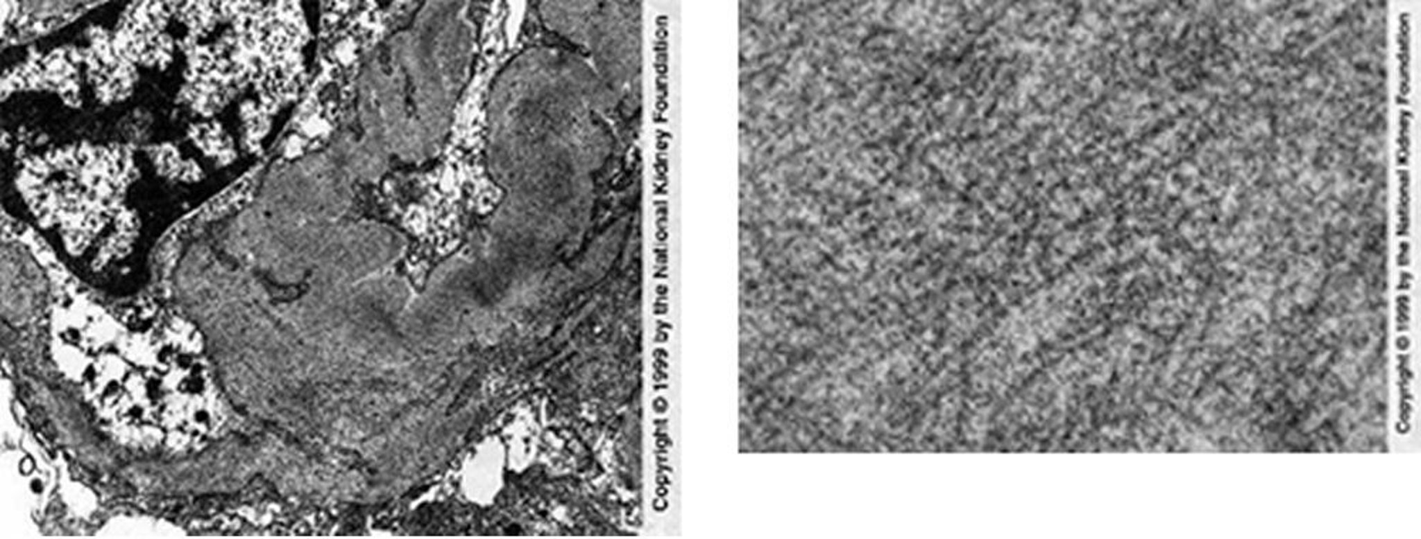
Фібрилярна гломерулопатія (фібрили)
Randomly arranged fibrils in mesangial and capillary loops are seen on transmission electron microscopy (left). Negative Congo red stains are necessary to exclude renal amyloidosis (×25,625). The right image shows a high-power view of fibrils, which are coarser in diameter than amyloid deposits (×98,000).
Image provided by Agnes Fogo, MD, and the American Journal of Kidney Diseases' Atlas of Renal Pathology (see www.ajkd.org).
Серед цих тем