


Nefrite tubulointersticial aguda

Nefrite tubulointersticial aguda
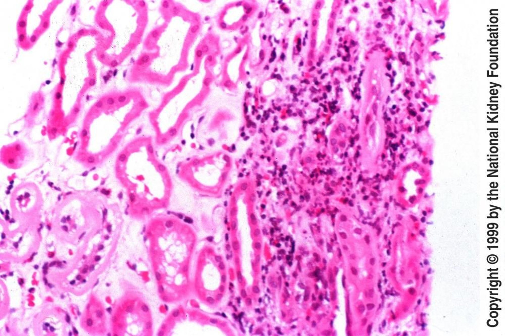
Amostra de biópsia na nefrite tubulointersticial aguda demonstra edema intersticial com infiltrado de eosinófilos, linfócitos e plasmócitos (coloração hematoxilina-eosina, × 200).
Imagem fornecida por Agnes Fogo, MD, e the American Journal of Kidney Diseases' Atlas of Renal Pathology (ver www.ajkd.org).
Nesses tópicos