

Temporal bone fractures can occur after severe blunt trauma to the head and sometimes involve structures of the ear, causing hearing loss, vertigo, balance disturbance, or facial nerve paralysis.
Temporal bone fractures are suggested by the following physical findings:
Battle sign (postauricular ecchymosis)
Bleeding in the external auditory canal
Bleeding may come from the middle ear (hemotympanum) through a ruptured tympanic membrane or from a fracture line in the ear canal. Hemotympanum makes the tympanic membrane appear blue-black. Cerebrospinal fluid otorrhea indicates a communication between the middle ear and the subarachnoid space.
Temporal bone fractures are classified as either otic capsule-sparing (90%) or otic capsule-violating (10%) (1). Otic capsule-sparing fractures do not involve the bony labyrinth (cochlea, vestibule, or semicircular canals), while otic capsule-violating fractures extend through one or more of these structures. This classification system has better predictive ability for sensorineural hearing loss (incidence is 5 to 20% in otic capsule sparing versus 40 to 100% in otic capsule-violating [2, 3]), vestibular dysfunction, and facial nerve paralysis (incidence is approximately 45 to 70% in otic capsule sparing versus 6 to 12% in otic capsule-violating [1, 2, 3]) than the previously used system that described longitudinal versus transverse fractures.
Temporal bone fracture can also cause conductive hearing loss if injury to the ossicular chain occurs.
Rarely, fluctuating sensorineural hearing loss and vestibular dysfunction occur with temporal bone fracture and may be due to a perilymph fistula. Immediate complete facial paralysis may indicate a severed or crushed facial nerve, whereas delayed-onset complete facial paralysis usually indicates edema within an intact nerve.

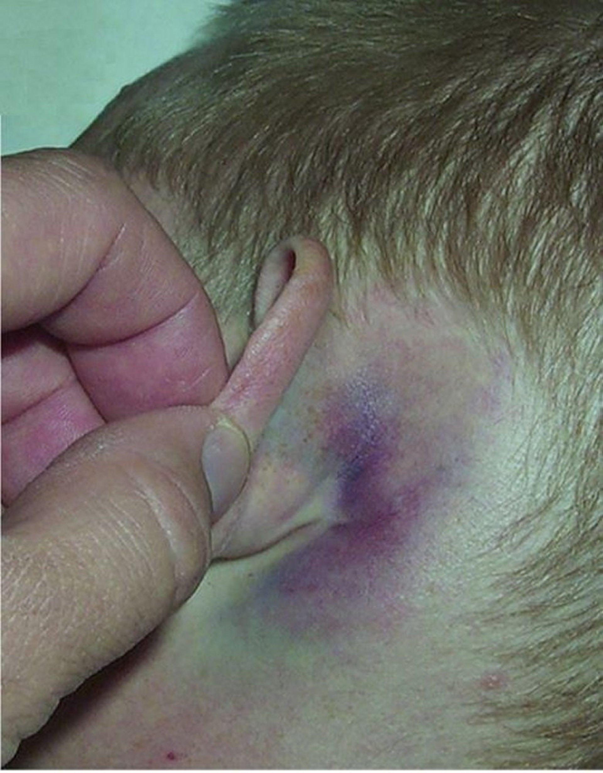
This photo shows the Battle sign (mastoid ecchymosis).
© Springer Science+Business Media
General references
1. Little SC, Kesser BW. Radiographic classification of temporal bone fractures: clinical predictability using a new system. Arch Otolaryngol Head Neck Surg. 2006;132(12):1300-1304. doi:10.1001/archotol.132.12.1300
2. Park E, Chang YS, Kim BJ, et al. Improved Prediction of Hearing Loss after Temporal Bone Fracture by Applying a Detailed Classification for Otic Capsule-Violating Fracture: A Wide Scope Analysis with Large Case Series. Otol Neurotol. 2023;44(2):153-160. doi:10.1097/MAO.0000000000003786
3. Dunklebarger J, Branstetter B 4th, Lincoln A, et al. Pediatric temporal bone fractures: current trends and comparison of classification schemes. Laryngoscope. 2014;124(3):781-784. doi:10.1002/lary.21891
Diagnosis of Temporal Bone Fractures
CT
Assessment of hearing and facial nerve function
If a temporal bone fracture is suspected, immediate CT of the head that includes high resolution CT of the temporal bone is recommended.
The Weber and Rinne tuning fork tests can be done during the initial physical examination in conscious patients to help differentiate between conductive and sensorineural hearing loss. However, formal audiometric examination is required for all patients with temporal bone fractures.
If facial paralysis is present, electrical testing of the facial nerve is warranted.
Treatment of Temporal Bone Fractures
Management of facial paralysis, hearing loss, vestibular dysfunction, and cerebrospinal fluid (CSF) leakage
Treatment is based on symptoms and complications, including hearing loss, vestibular dysfunction, facial nerve injury, or CSF leakage (1).
Facial paralysis is managed initially with observation. Surgical exploration may be considered for immediate complete paralysis with unfavorable electrodiagnostic findings and imaging showing displaced fracture through the fallopian canal.
Conductive hearing loss is managed initially with observation, and often resolves spontaneously. If hearing loss is persistent, CT should be performed to evaluate for ossicular injury, and ossiculoplasty may be required (2).
Sensorineural hearing loss after otic capsule-sparing temporal bone fracture often improves spontaneously (1). For persistent hearing loss in either type of fracture, hearing aids or cochlear implantation may be required. However, in the rare case of fluctuating sensorineural hearing loss, an exploratory tympanotomy to search for a perilymph fistula may be indicated.
There are no high-quality data regarding use of glucocorticoids as treatment of hearing loss following temporal bone fracture.
Vestibular dysfunction may resolve spontaneously. Persistent symptoms are managed with vestibular rehabilitation to improve balance and gaze stability. If the cause is perilymph fistula, repair may reduce severity and frequency of vertiginous episodes.
Patients with CSF otorrhea should be observed and hospitalization may be appropriate. The ear canal should not be irrigated or manipulated. Meningitis is a risk and patients should be instructed to report fever, new or worsening severe headache, photophobia, nuchal rigidity, confusion or altered mental status, focal neurologic deficits, or persistent, high-volume CSF drainage (3). The leak usually resolves spontaneously within a few days, although a lumbar drain or surgical closure of the defect is occasionally required.
Treatment references
1. Diaz RC, Cervenka B, Brodie HA. Treatment of Temporal Bone Fractures. J Neurol Surg B Skull Base. 2016;77(5):419-429. doi:10.1055/s-0036-1584197
2. Tu A, Doerfer KW. Ossiculoplasty for Trauma. Otolaryngol Clin North Am. Published online January 10, 2026. doi:10.1016/j.otc.2025.11.005
3. Nasrollahi TS, Kuan EC. Conservative Management of Cerebrospinal Fluid Leaks. Otolaryngol Clin North Am. Published online January 22, 2026. doi:10.1016/j.otc.2025.12.001
Key Points
Temporal bone fracture can cause blood coming from the ear, blood behind the tympanic membrane, hearing loss, vestibular dysfunction, and/or facial nerve paralysis.
Perform CT with attention to the temporal bone, refer patients for audiometry, and if facial nerve paralysis is suspected, arrange electrical testing of the facial nerve.
Direct treatment toward management of facial nerve injury, hearing loss, vestibular dysfunction, and cerebrospinal fluid leakage.


