

Keratoacanthomas are round, firm, usually flesh-colored nodules with sharply sloping borders and a characteristic central crater containing keratinous material; they usually resolve spontaneously, but some may be a well-differentiated form of squamous cell carcinoma. Diagnosis is by biopsy or excision. Treatment is with surgery or injections of methotrexate or 5-fluorouracil.
Keratoacanthomas are rapidly growing, dome-shaped skin tumors with a central keratin-filled crater. They most commonly arise on sun-exposed areas in older adults (1). There is a male predilection. (See also Overview of Skin Cancer.)

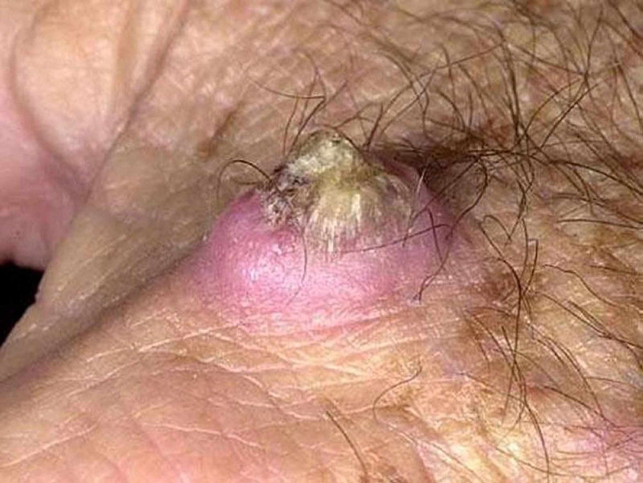
A keratoacanthoma is a round, firm nodule with sharply sloping borders and a characteristic central crater.
Photo provided by Thomas Habif, MD.

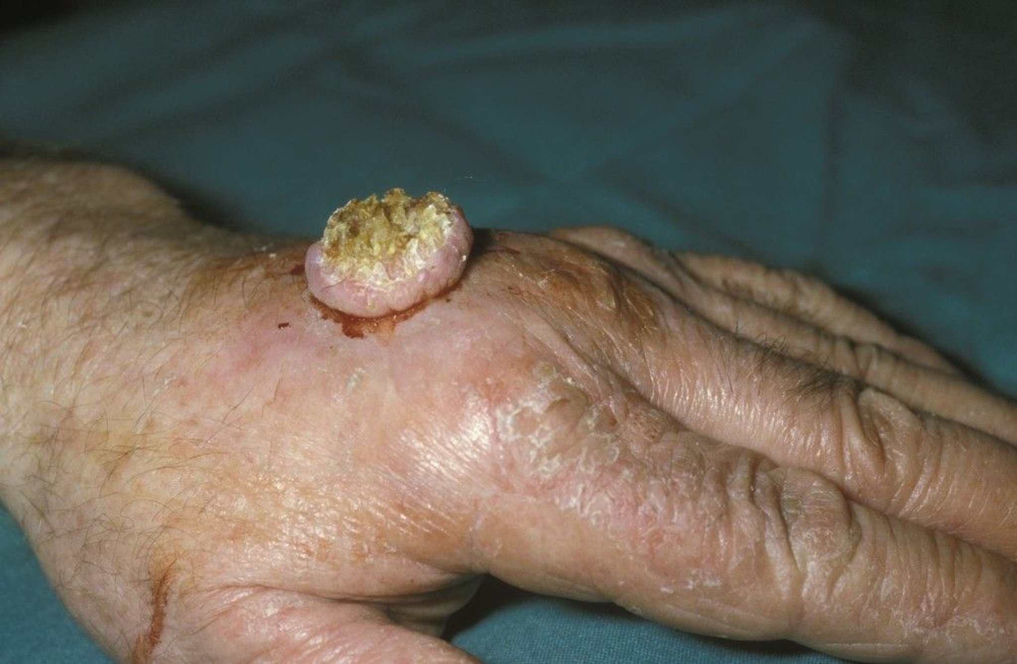
This photo shows a large keratoacanthoma on the hand of a patient.
DR M.A. ANSARY/SCIENCE PHOTO LIBRARY
General reference
1. Kwiek B, Schwartz RA. Keratoacanthoma (KA): An update and review. J Am Acad Dermatol.2016 Jun;74(6):1220-33. doi: 10.1016/j.jaad.2015.11.033. Epub 2016 Feb 4. PMID: 26853179.
Etiology of Keratoacanthoma
Historically, keratoacanthomas have been considered a variant of cutaneous squamous cell carcinomas (1). Opinion is divided on whether keratoacanthomas are a benign lesion, a variant of well-differentiated squamous cell carcinoma (SCC), or a borderline entity, as they share clinical and histopathologic features with squamous cell carcinoma and in rare cases, may be aggressive.
Unlike most squamous cell carcinomas, keratoacanthomas have a tendency to regress or involute. They may also have a different genetic basis involving activation of apoptotic pathways (2).
Etiology references
1. Tisack A, Fotouhi A, Fidai C, et al. A clinical and biological review of keratoacanthoma. Br J Dermatol. 2021 Sep;185(3):487-498. doi: 10.1111/bjd.20389. Epub 2021 Jun 14. PMID: 33864244.
2. Nirenberg A, Steinman H, Dixon A. Keratoacanthoma: Update on the Debate. Am J Dermatopathol. 2021 Apr 1;43(4):305-307. doi: 10.1097/DAD.0000000000001872. Erratum in: Am J Dermatopathol. 2021 Dec 1;43(12):1006. doi: 10.1097/DAD.0000000000001983. PMID: 33395044.
Symptoms and Signs of Keratoacanthoma
Development is rapid. Usually the lesion reaches its full size, typically 1 to 3 cm but sometimes > 5 cm, within 1 or 2 months. Common sites are sun-exposed areas: the face, the forearms, and the dorsum of the hands.
Spontaneous involution may start within a few months, but involution does not occur universally.
Diagnosis of Keratoacanthoma
Biopsy or excision
Because this lesion cannot be relied on to involute spontaneously, biopsy or excision is recommended.
Treatment of Keratoacanthoma
Surgery or injections of methotrexate or 5-fluorouracilSurgery or injections of methotrexate or 5-fluorouracil
Spontaneous involution may leave substantial scarring; surgery or intralesional injections with methotrexate or 5-fluorouracil usually yield better cosmetic results, and excision allows histologic confirmation of the diagnosis. Spontaneous involution may leave substantial scarring; surgery or intralesional injections with methotrexate or 5-fluorouracil usually yield better cosmetic results, and excision allows histologic confirmation of the diagnosis.
Surgical excision is associated with a very high cure rate and low risk of recurrence (1).
Treatment reference
1. Moss M, Weber E, Hoverson K, et al. Management of Keratoacanthoma: 157 Tumors Treated With Surgery or Intralesional Methotrexate. Dermatol Surg. 2019 Jul;45(7):877-883. doi: 10.1097/DSS.0000000000001739. PMID: 30608293.
Prevention of Keratoacanthoma
Because keratoacanthoma risk may increase with increasing ultraviolet (UV) radiation exposure, a number of measures are often recommended to limit exposure (eg, sun avoidance measures, use of protective clothing, use of sunscreen). For more detailed information, see Prevention of Effects of Sun Exposure.
Drug Information for the Topic


