


Noncancerous (benign) and cancerous (malignant) tumors can affect the nail unit, causing a changes in nail texture and/or color (dystrophy). Many of these are tumors that originate in tissues around but not in the nail.
Noncancerous tumors include myxoid cysts (benign, fluid-filled swellings), pyogenic granulomas, and glomus tumors.
Cancerous tumors include Bowen disease (an early form of skin cancer), squamous cell carcinoma, and malignant melanoma. When doctors suspect cancer, they do a biopsy and may recommend complete removal of the tumor as soon as possible.

The arrow points to a myxoid cyst. Myxoid cysts typically develop close to the fingernail or toenail, often in people with osteoarthritis.
© Springer Science+Business Media
A dark band in the nail may be the initial sign of malignant melanoma of the nail. Pigment cells of the nail-making tissue known as the nail matrix may become malignant and develop into a melanoma. A worrisome sign is known as Hutchinson sign. The Hutchinson sign is black, brown, or gray discoloration that extends to the area around the nail, such as the cuticle or nail fold (the fold of hard skin at the sides of the nail plate where the nail and the skin meet). This sign may mean there is melanoma in the nail bed (the soft tissue underneath the nail plate that attaches the nail to the finger). When this sign is present, doctors do a biopsy of the nail and the affected skin. Melanoma may also occur without the Hutchinson sign. Melanoma occurs mostly in adults and is very rare in children.

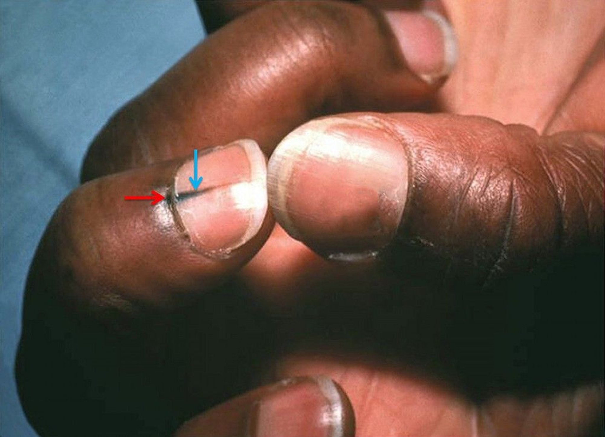
In this photo, the middle finger is marked by lengthwise discoloration of the nail (melanonychia striata; blue arrow) and hyperpigmentation that extends across the half-moon at the base of the nail (lunula) to the cuticle (Hutchinson sign; red arrow).
Image courtesy of Carl Washington, MD, and Mona Saraiya, MD, MPH, via the Public Health Image Library of the Centers for Disease Control and Prevention.


