

Sesamoiditis is pain at the sesamoid bones beneath the head of the first metatarsal, with or without inflammation or fracture. Diagnosis is usually clinical. Treatment is usually modification of footwear and orthotics.

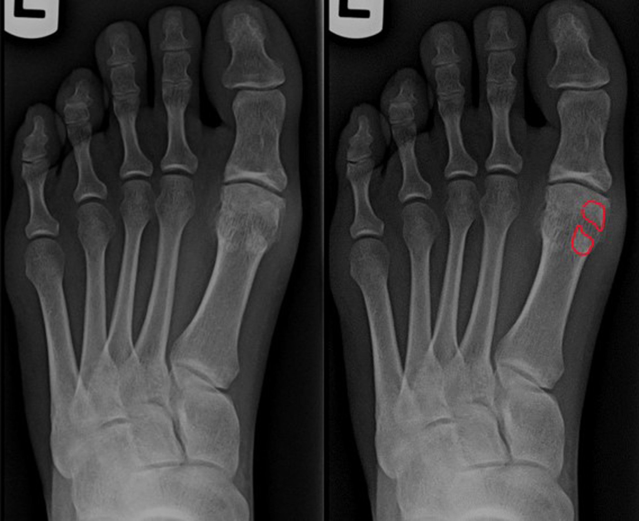
Anteroposterior view of the left foot shows a tibial sesamoid fracture. Note increased diastasis between the fracture fragments and the jagged fracture lines on the highlighted view. Differentiation of a bipartite sesamoid is important to ensure proper patient care.
Image courtesy of James C. Connors, DPM.
Sesamoiditis is a common cause of metatarsalgia. The 2 semilunar-shaped sesamoid bones are located within the flexor hallucis brevis tendon and aid the foot in locomotion. The medial bone is the tibial sesamoid, and the lateral bone is the fibular sesamoid. Direct trauma or positional change of the sesamoids due to alterations in foot structure (eg, lateral displacement of a sesamoid due to lateral deviation of the great toe) can make the sesamoids painful. Sesamoiditis is particularly common among dancers, joggers, and people who have high-arched feet or wear high heels. Many people with bunions have tibial sesamoiditis. This can sometimes be confused clinically with gout.
(See also Overview of Foot and Ankle Disorders.)
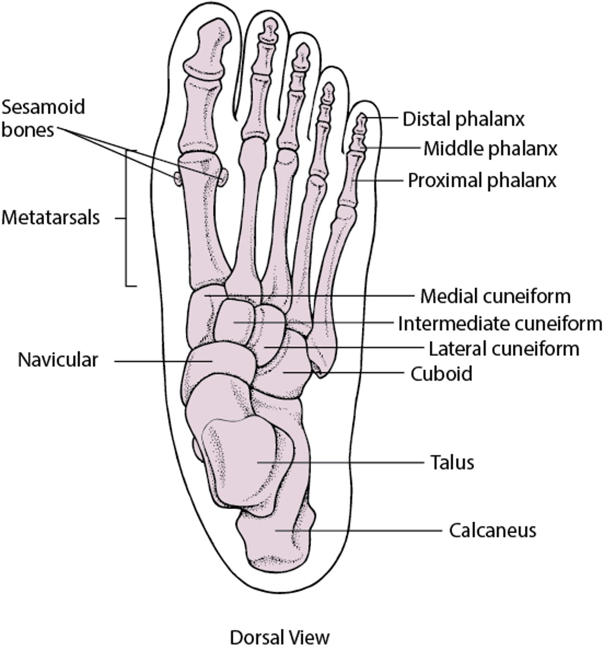
Bones of the Foot

Symptoms and Signs of Sesamoiditis
The pain of sesamoiditis is beneath the head of the first metatarsal; the pain is usually made worse by ambulation and may be worse when wearing flexible thin-soled or high-heeled shoes. Occasionally, inflammation occurs, causing mild warmth and swelling or occasionally redness that may extend medially and appear to involve the first metatarsophalangeal joint. Sesamoid fracture can also cause pain, moderate swelling, and possibly inflammation.
Diagnosis of Sesamoiditis
History and physical examination
Arthrocentesis if there is circumferential joint swelling
Imaging if fracture, osteoarthritis, or displacement is suspected
With the foot and first (great) toe dorsiflexed, the examiner inspects the metatarsal head and palpates each sesamoid. Tenderness is localized to a sesamoid, usually the tibial sesamoid. Hyperkeratotic tissue may indicate that a wart or discrete callus is causing pain.
If inflammation causes circumferential swelling around the first metatarsophalangeal joint, arthrocentesis is usually indicated to exclude gout and infectious arthritis.
If fracture, osteoarthritis, or displacement is suspected,radiographs are taken. Sesamoids separated by cartilage or fibrous tissue (bipartite sesamoids) may appear fractured on radiographs. If radiographs are equivocal, MRI is indicated for diagnosis.

Treatment of Sesamoiditis
New shoes, orthotics, or both
In patients with sesamoiditis, simply not wearing shoes that cause pain may be sufficient. If symptoms of sesamoiditis persist, offloading pads as well as shoes with a thick sole and orthotics are prescribed and help by reducing sesamoid pressure. If fracture without displacement is present, conservative therapy may be sufficient and may also involve immobilization of the joint with the use of a flat, rigid, surgical shoe.
Nonsteroidal anti-inflammatory drugs (NSAIDs) and injections of a glucocorticoid combined with a local anesthetic solution can be helpful (see Considerations for Using Glucocorticoid Injections).
Although surgical removal of the sesamoid may help in recalcitrant cases, it is controversial because of the potential for disturbing biomechanics and mobility of the foot.
Key Points
Dancers, joggers, and people who have high-arched feet, wear high heels, or have bunions can develop pain at the sesamoids beneath the head of the first metatarsal.
Pain is worse when weight-bearing, particularly when wearing certain shoes.
Diagnose based on clinical findings; exclude infection and gout with synovial fluid analysis when swelling is present and exclude suspected fracture with radiographs and sometimes MRI.
Prescribe new, thick-soled shoes, offloading pads, and orthotics that decrease pressure on the sesamoids, or both.


