

Erosive gastritis is gastric mucosal erosion caused by damage to mucosal defenses. It is typically acute, manifesting with bleeding, but may be subacute or chronic with few or no symptoms. Diagnosis is by endoscopy. Treatment is supportive, with removal of the inciting cause and initiation of acid-suppressant therapy. Certain patients being treated in an intensive care unit (eg, those with head trauma, burn, multisystem trauma, or who are being mechanically ventilated) benefit from prophylaxis with acid suppressants.
(See also Overview of Acid Secretion and Overview of Gastritis.)
Common causes of erosive gastritis include
Nonsteroidal anti-inflammatory drugs (NSAIDs)
Alcohol
Stress
Less common causes of erosive gastritis include
Radiation
Viral infection (eg, cytomegalovirus)
Vascular injury
Direct trauma (eg, nasogastric tubes)
Superficial erosions and punctate mucosal lesions occur. These may develop as soon as 12 hours after the initial insult. Deep erosions, ulcers, and sometimes perforation may occur in severe or untreated cases. Lesions typically occur in the body, but the antrum may also be involved.
Acute stress gastritis, a form of erosive gastritis, can occur in critically ill patients. The incidence increases with duration of intensive care unit stay and length of time the patient is not receiving enteral feeding. Pathogenesis likely involves hypoperfusion of the gastrointestinal mucosa, resulting in impaired mucosal defenses. Patients with head injury or burns may also have increased secretion of acid.
Symptoms and Signs of Erosive Gastritis
Patients with mild erosive gastritis are often asymptomatic, although some complain of dyspepsia, nausea, or vomiting.
Often, the first sign is hematemesis, melena, or blood in the nasogastric aspirate, usually within 2 to 5 days of the inciting event. Bleeding is usually mild to moderate, although it can be massive if deep ulceration is present, particularly in acute stress gastritis.
Diagnosis of Erosive Gastritis
Endoscopy
Acute and chronic erosive gastritis are diagnosed endoscopically.

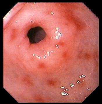
This photo shows eroded and erythematous areas in the stomach lining resulting from prolonged nonsteroidal anti-inflammatory drug use.
Treatment of Erosive Gastritis
For bleeding: Endoscopic hemostasis
For acid suppression: A proton pump inhibitor or histamine-2 receptor antagonist
In severe gastritis, bleeding is managed with IV fluids and blood transfusion as needed. Endoscopic hemostasis should be attempted, with surgery a fallback procedure if bleeding cannot be controlled endoscopically. Angiography is unlikely to stop severe gastric bleeding because of the many collateral vessels supplying the stomach. Acid-suppressing therapy should be started if the patient is not already receiving it.
For milder gastritis, removing the offending agent and using medications to reduce gastric acidity (see Medications for the Treatment of Gastric Acidity) to limit further injury and promote healing may be all that is required.
Prevention of Erosive Gastritis
Prophylaxis with acid-suppressive medications can reduce the incidence of acute stress gastritis. However, it mainly benefits certain patients who are at high risk of bleeding and being treated in an intensive care unit, including those with severe burns, central nervous system trauma, coagulopathy, sepsis, shock, multiple trauma, mechanical ventilation for > 48 hours, chronic liver disease, acute kidney injury, hepatic or renal failure, multiorgan dysfunction, and history of peptic ulcer or gastrointestinal bleeding.
One guideline recommends stress ulcer prophylaxis with histamine-2 receptor antagonists or proton pump inhibitors in critically ill adults with coagulopathy, shock, or chronic liver disease who are being treated in an ICU (1).
There is a possible increased risk of nosocomial pneumonia in critically ill patients receiving acid suppression. A recent meta-analysis concluded that proton pump inhibitors (PPIs) and histamine-2 receptor antagonists may increase the risk of pneumonia (absolute increases 5% for PPIs and 3.4% for histamine-2 receptor antagonists; 2). However, a previous large clinical study of a PPI for patients at risk of gastrointestinal bleeding in the intensive care unit found no increased incidence of pneumonia (3).
Early enteral feeding also can decrease the incidence of bleeding.
Acid suppression is not recommended for patients simply taking nonsteroidal anti-inflammatory drugs unless they have previously had an ulcer.
Prevention references
1. MacLaren R, Dionne JC, Granholm A, et al: Society of Critical Care Medicine and American Society of Health-System Pharmacists Guideline for the Prevention of Stress-Related Gastrointestinal Bleeding in Critically Ill Adults. Crit Care Med 52(8):e421–e430, 2024. doi:10.1097/CCM.0000000000006330
2. Wang Y, Ye Z, Ge L, et al: Efficacy and safety of gastrointestinal bleeding prophylaxis in critically ill patients: Systematic review and network meta-analysis. BMJ 368:l6744, 2020. doi: 10.1136/bmj.l6744PMCID
3. Krag M, Marker S, Perner A, et al: Pantoprazole in patients at risk for gastrointestinal bleeding in the ICU. N Engl J Med 379(23):2199–2208, 2018. doi: 10.1056/NEJMoa1714919
Key Points
Erosive gastritis is erosion of the gastric mucosa due to damage to the gastric mucosal barrier.
Common causes include nonsteroidal anti-inflammatory drugs (NSAIDs), alcohol, and stress; acute stress gastritis occurs in about 5% of critically ill patients.
Symptoms include dyspepsia, nausea, and vomiting, but mild cases may be asymptomatic.
Gastrointestinal bleeding (hematemesis or melena) may be the initial sign.
Diagnose with upper endoscopy.
Treat with a proton pump inhibitor (PPI) or histamine-2 receptor antagonist and removal of the causative agent; treat bleeding patients with IV fluids and/or blood transfusion as needed and endoscopic hemostasis with surgical backup.
Prevention of acute stress gastritis with a PPI is recommended for selected critically ill patients, though this may slightly increase the risk of pneumonia.
Prevention of NSAID-related gastritis with a PPI or histamine-2 receptor antagonists is not indicated unless there is a prior history of peptic ulcer disease.


