

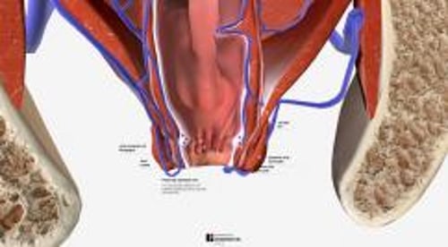
The anal canal begins at the anal verge and ends at the anorectal junction (pectinate line, mucocutaneous junction, dentate line), where there are 8 to 12 anal crypts and 5 to 8 papillae. The canal is lined with anoderm, a continuation of the external skin. The anal canal and adjacent skin are innervated by somatic sensory nerves and are highly susceptible to painful stimuli.
Venous drainage from the anal canal occurs through the caval system, but the anorectal junction can drain into both the portal and caval systems. Lymphatics from the anal canal pass to the internal iliac nodes, the posterior vaginal wall, and the inguinal nodes. The venous and lymphatic distributions determine how malignant disease and infection spread.
The rectum is a continuation of the sigmoid colon beginning at the level of the 3rd sacral vertebra and continuing to the anorectal junction. The rectal lining consists of red, glistening glandular mucosa, which has an autonomic nerve supply and is relatively insensitive to pain.
Venous drainage occurs through the portal system. Lymphatic return from the rectum occurs along the superior hemorrhoidal vascular pedicle to the inferior mesenteric and aortic nodes.
The sphincteric ring encircling the anal canal is composed of the internal sphincter, the central portion of the levators, and components of the external sphincter. Anteriorly, it is more vulnerable to trauma, which can result in incontinence. The puborectalis forms a muscular sling around the rectum for support and assistance in urination and defecation.
Anorectal disorders include
(See also Overview of Foreign Bodies of the Gastrointestinal Tract.)
History
History should include the details of bleeding, pain, protrusion, discharge, swelling, abnormal sensations, bowel movements, incontinence, stool characteristics, use of cathartics and enemas, and abdominal and urinary symptoms.
All patients should be asked about anal intercourse and other possible causes of trauma and infection.
Physical examination
Examination should be done gently and with good lighting. It consists of external inspection, perianal and intrarectal digital palpation, abdominal examination, and rectovaginal bidigital palpation.
Anoscopy and rigid or flexible sigmoidoscopy to 15 to 60 cm above the anal verge are often included (see also Anoscopy and Sigmoidoscopy). Inspection, palpation, and anoscopy and sigmoidoscopy are best done with the patient in the left lateral position (Sims position) or inverted on a tilt table. In cases of painful anal lesions, topical (lidocaine 5% ointment), regional, or even general anesthesia may be required. If it can be tolerated, a cleansing phosphate enema may facilitate sigmoidoscopy. Samples may be taken for biopsies, smears, and cultures, and imaging studies are done if indicated.). Inspection, palpation, and anoscopy and sigmoidoscopy are best done with the patient in the left lateral position (Sims position) or inverted on a tilt table. In cases of painful anal lesions, topical (lidocaine 5% ointment), regional, or even general anesthesia may be required. If it can be tolerated, a cleansing phosphate enema may facilitate sigmoidoscopy. Samples may be taken for biopsies, smears, and cultures, and imaging studies are done if indicated.


