

Angiography is sometimes called conventional angiography to distinguish it from CT angiography (CTA) and magnetic resonance angiography (MRA). Angiography provides detailed images of blood vessels, commonly those in the heart, lungs, brain, and legs. Angiography can provide still images or motion pictures (called cineangiography).
IV contrast is injected through a catheter inserted into a blood vessel that connects with the vessel to be imaged. A local anesthetic or a sedative may be used. If the catheter is inserted into an artery, the insertion site must be steadily compressed for 10 to 20 minutes after all instruments are removed to reduce the risk of bleeding at the puncture site.
Angiography, although invasive, is relatively safe.
Uses of Angiography
CTA and MRA are often done instead of conventional angiography. However, conventional angiography is the traditional gold standard for evaluating vascular lesions (eg, stenosis, obstruction, arteriovenous or other vascular malformations, aneurysms, dissections, vasculitis).
Conventional angiography is usually done before therapeutic angiographic procedures such as angioplasty, vascular stenting, and embolization of tumors and vascular malformations.
Common uses of conventional angiography include the following:
Coronary angiography is usually done before percutaneous or surgical interventions involving the coronary arteries or heart valves. It is usually done with cardiac catheterization.
Cerebral angiography may be indicated after stroke or transient ischemic attack (TIA)—eg, if stent placement or carotid endarterectomy is being considered.
Iliac and femoral angiography may be indicated before interventions to treat peripheral arterial disease.
Aortography is sometimes done to diagnose and provide anatomic detail about aortic aneurysms, aortic dissection, and aortic regurgitation.
Angiography of the retinal arteries can be done using fluorescein dye.can be done using fluorescein dye.
Conventional pulmonary angiography, at one time the gold standard for diagnosis of pulmonary embolism, has largely been replaced by CT pulmonary angiography (CTPA), which is less invasive.
Variations of Angiography
Digital subtraction angiography
Images of blood vessels are taken before and after contrast injection; then a computer subtracts the pre-contrast image from the post-contrast image. Images of extraneous structures are thus eliminated, isolating images of the blood vessel lumens opacified by contrast.

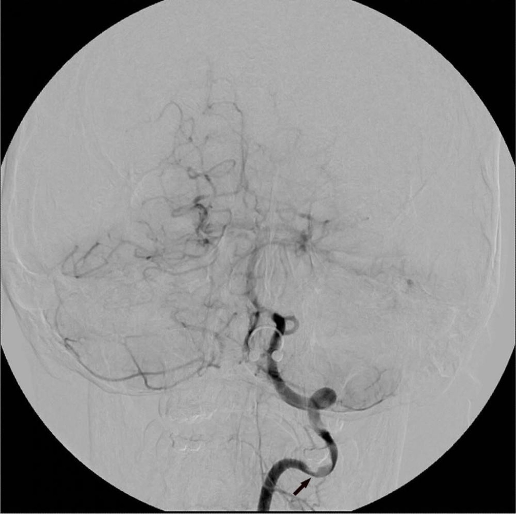
Digital subtraction angiography (DSA) of the left vertebral artery shows a focal area of stenosis (arrow).
Image courtesy of Hakan Ilaslan, MD.
Disadvantages of Angiography
Contrast reactions occasionally occur.
The injection site may bleed if the injected blood vessel ruptures; a painful hematoma can form. Rarely, the site becomes infected; it becomes red and swollen and exudes a purulent discharge within a few days after the injection.
Rarely, an artery is injured by the catheter, or an atherosclerotic plaque dislodges, causing an embolism distally. Very rarely, shock, seizures, acute kidney injury, and cardiac arrest occur.
Risk of complications is higher in older patients, although it is still low.
The radiation dose used in angiography can vary and be significant (eg, coronary angiography is associated with an effective radiation dose of 4.6 to 15.8 mSv).
Angiography must be done by highly skilled physicians, usually specially trained interventional radiologists or cardiologists.
Drug Information for the Topic


