


Fibrillary Glomerulopathy (Fibrils)

Fibrillary Glomerulopathy (Fibrils)
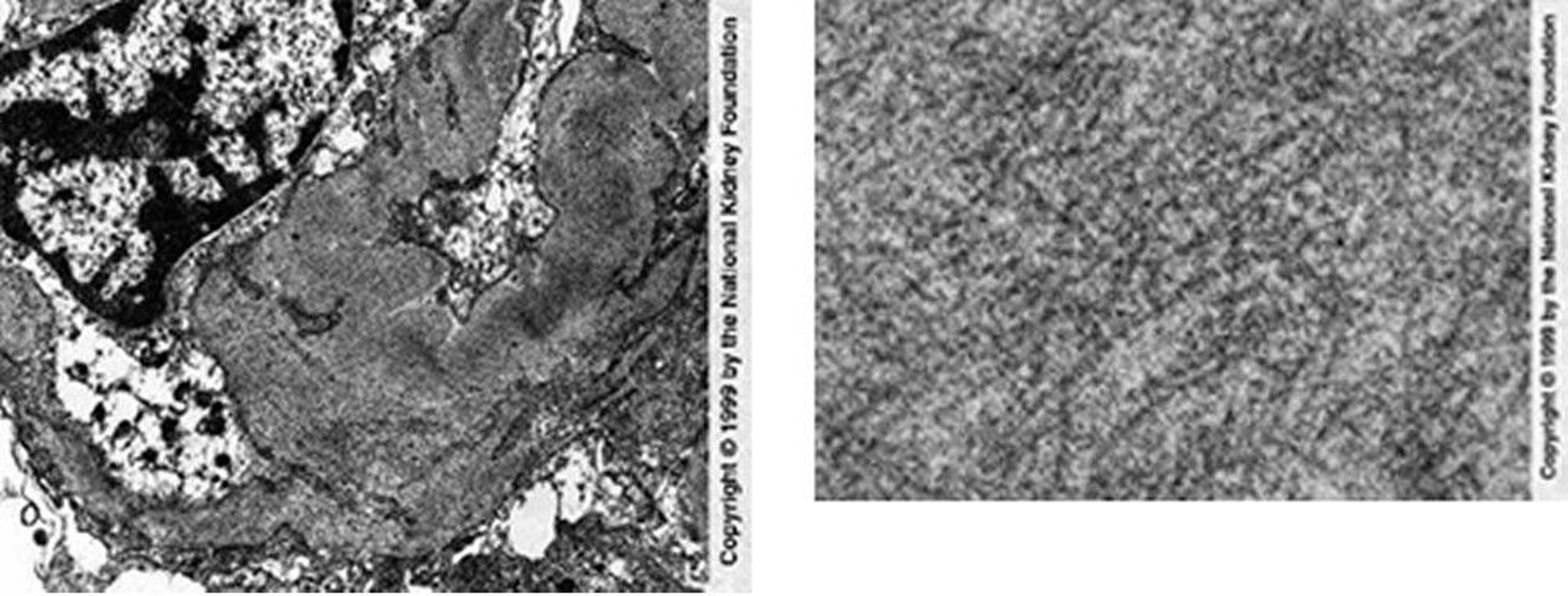
Randomly arranged fibrils in mesangial and capillary loops are seen on transmission electron microscopy (left). Negative Congo red stains are necessary to exclude renal amyloidosis (×25,625). The right image shows a high-power view of fibrils, which are coarser in diameter than amyloid deposits (×98,000).
Image provided by Agnes Fogo, MD, and the American Journal of Kidney Diseases' Atlas of Renal Pathology (see www.ajkd.org).
In these topics