


The peripheral nervous system consists of more than 100 billion nerve cells (neurons) that run throughout the body like strings, making connections with the brain, other parts of the body, and often with each other.
Peripheral nerves consist of bundles of nerve fibers. These fibers are wrapped with many layers of tissue composed of a fatty substance called myelin. These layers form the myelin sheath, which speeds the conduction of nerve impulses along the nerve fiber. Nerves conduct impulses at different speeds depending on their diameter and on the amount of myelin around them.
The peripheral nervous system has 2 parts:
The somatic nervous system
The autonomic nervous system
Somatic nervous system
The somatic nervous system consists of nerves that connect the brain and spinal cord with muscles controlled by conscious effort (voluntary or skeletal muscles) and with sensory receptors in the skin. Sensory receptors are specialized endings of nerve fibers that detect information in and around the body.
Autonomic nervous system
The autonomic system connects the brain stem and spinal cord with internal organs and regulates internal body processes that require no conscious effort and that people are thus usually unaware of (see Overview of the Autonomic Nervous System). Examples are the rate and strength of heart contractions, blood pressure, the rate of breathing, and the speed at which food passes through the digestive tract.
The autonomic nervous system has 2 divisions:
Sympathetic division: Its main function is to prepare the body for stressful or emergency situations—for "fight or flight."
Parasympathetic division: Its main function is to maintain normal body functions during ordinary situations.
These divisions work together, usually with one activating and the other inhibiting the actions of internal organs. For example, the sympathetic division increases pulse, blood pressure, and breathing rates, and the parasympathetic system decreases each of them.
Typical Structure of a Nerve Cell
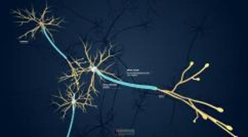
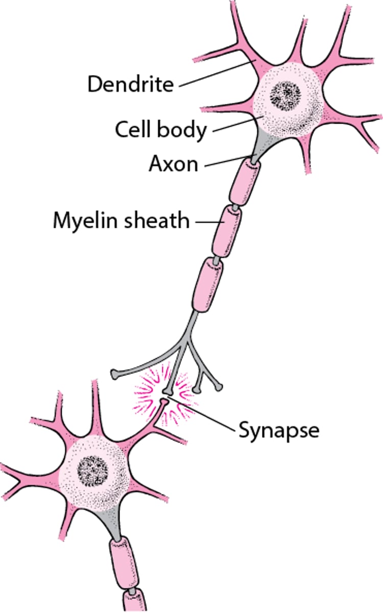
A nerve cell (neuron) consists of a large cell body and nerve fibers—one elongated extension (axon) for sending impulses and usually multiple branches (dendrites) for receiving impulses. Impulses from the axon of 1 nerve cell cross a synapse (the junction between 2 nerve cells) to the dendrite of another cell. Each large axon is surrounded by oligodendrocytes in the brain and spinal cord and by Schwann cells in the peripheral nervous system. The membranes of these cells consist of a fat (lipoprotein) called myelin. The membranes are wrapped tightly around the axon, forming a multilayered sheath. This myelin sheath resembles insulation, such as that around an electrical wire. Nerve impulses travel much faster in nerves with a myelin sheath than in those without one. |

Cranial nerves and spinal nerves
Cranial nerves directly connect the brain and the brain stem with the eyes, ears, nose, and throat and with various parts of the head, neck, and trunk. There are 12 pairs of cranial nerves and they transmit sensory information including touch, vision, taste, smell, and hearing. Cranial nerve II or the optic nerve, is actually not considered a peripheral nerve as it is an outcropping of the brain and is myelinated by oligodendrocytes, not Schwann cells (see Overview of the Cranial Nerves).
Spinal nerves connect the spinal cord with other parts of the body. The brain communicates with most of the body through the spinal nerves. There are 31 pairs of them, located at intervals along the length of the spinal cord (see Overview of Spinal Cord Disorders). Several cranial nerves and most spinal nerves are involved in both the somatic and autonomic parts of the peripheral nervous system.
Spinal nerves emerge from the spinal cord through spaces between the vertebrae. Each nerve emerges as 2 short branches (called spinal nerve roots): one at the front of the spinal cord and one at the back.
Motor nerve root (anterior nerve root): The motor root emerges from the front of the spinal cord. Motor nerve fibers carry commands from the brain and spinal cord to other parts of the body, particularly to skeletal muscles.
Sensory nerve root (posterior nerve root): The sensory root enters the back of the spinal cord. Sensory nerve fibers carry sensory information (about body position, light, touch, temperature, and pain) to the brain from other parts of the body. The sensory nerve fibers in each sensory nerve root carry information from a specific area of the body, called a dermatome.
After leaving the spinal cord, the corresponding motor and sensory nerve roots join to form a single spinal nerve.
Some of the spinal nerves form networks of interwoven nerves, called nerve plexuses. In a plexus, nerve fibers from different spinal nerves are sorted and recombined so that all fibers going to or coming from one area of a specific body part are put together into one nerve (see figure ).
There are 2 major nerve plexuses:
Brachial plexus: Sorts and recombines nerve fibers traveling to the arms and hands
Lumbosacral plexus: Sorts and recombines nerve fibers going to the legs and feet


