Tension pneumothorax is accumulation of air in the pleural space under pressure, compressing the lungs and decreasing venous return to the heart.
(See also Overview of Thoracic Trauma.)
Tension pneumothorax develops when a lung or chest wall injury is such that it allows air into the pleural space but not out of it (a one-way valve). As a result, air accumulates and compresses the lung, eventually shifting the mediastinum, compressing the contralateral lung, and increasing intrathoracic pressure enough to decrease venous return to the heart, causing shock. These effects can develop rapidly, particularly in patients undergoing positive pressure ventilation.

DU CANE MEDICAL IMAGING LTD/SCIENCE PHOTO LIBRARY
Causes include mechanical ventilation (most commonly) and simple (uncomplicated) pneumothorax with lung injury that fails to seal following penetrating or blunt chest trauma or failed central venous cannulation.
Symptoms and Signs of Tension Pneumothorax
Symptoms and signs initially are those of simple pneumothorax. As intrathoracic pressure increases, patients develop hypotension, tracheal deviation, and neck vein distention. The affected hemithorax is hyperresonant to percussion and often feels somewhat distended, tense, and poorly compressible to palpation.
Diagnosis of Tension Pneumothorax
History and physical examination
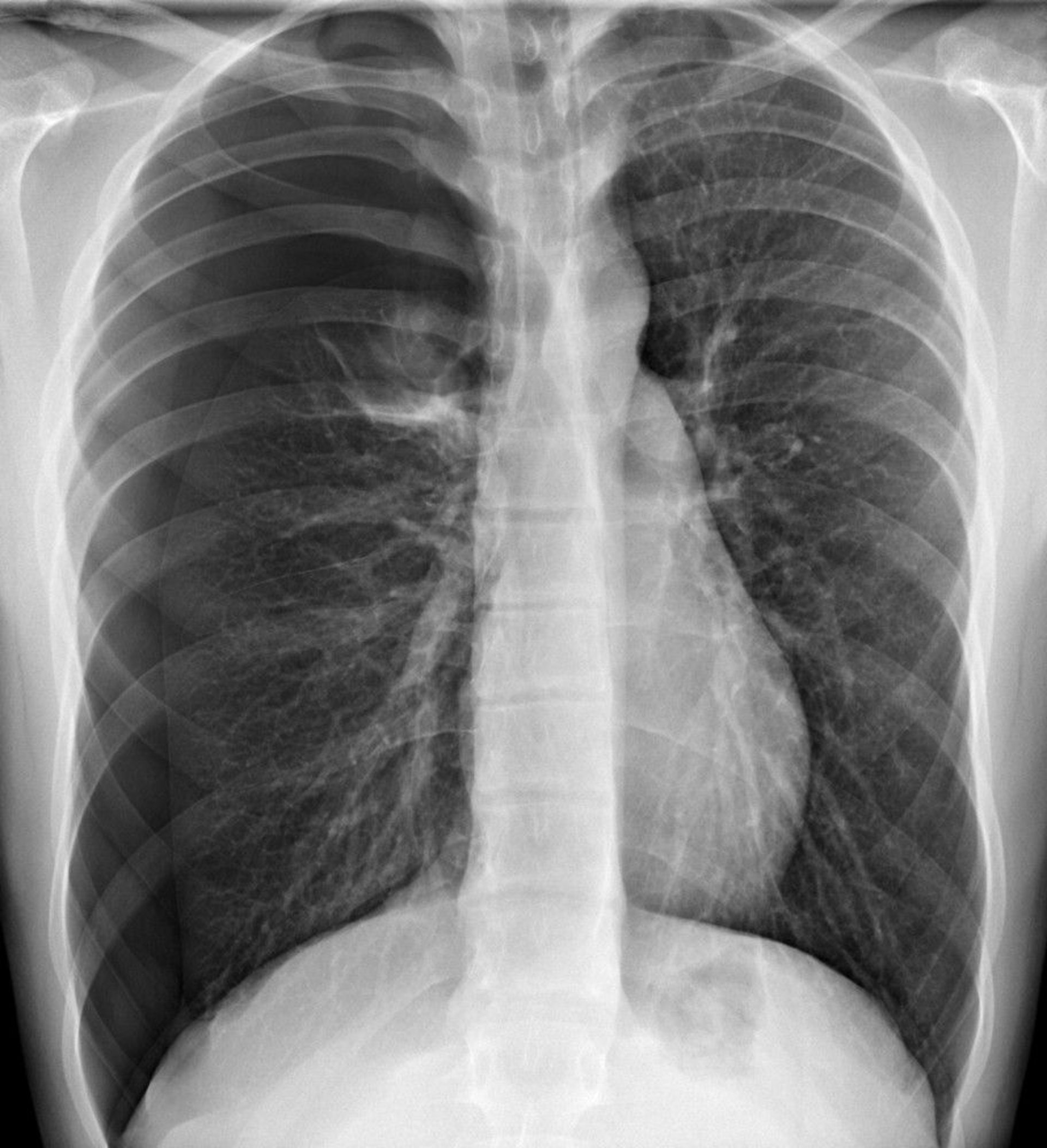
Tension pneumothorax should be diagnosed by clinical findings. Treatment should not be delayed pending radiographic confirmation. Although cardiac tamponade also can cause hypotension, neck vein distention, and sometimes respiratory distress, tension pneumothorax can be differentiated clinically by its unilateral absence of breath sounds and hyperresonance to percussion.
Pearls & Pitfalls
|
Treatment of Tension Pneumothorax
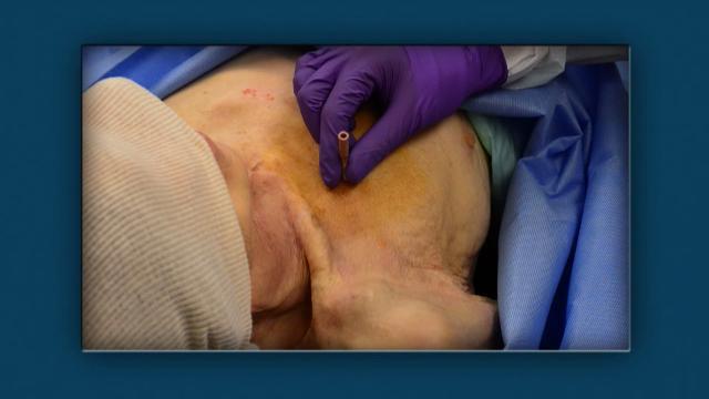
Needle decompression followed by tube thoracostomy
Treatment of tension pneumothorax is immediate needle decompression by inserting a large-bore (eg, 14- or 16-gauge) needle into the second intercostal space in the midclavicular line. The American College of Surgeons Advanced Trauma Life Support recommends placement in the fourth or fifth intercostal space along the midaxillary line, as it has been associated with improved success at decompression. Air will usually gush out. Because needle decompression causes a simple pneumothorax, tube thoracostomy should be done immediately thereafter.