

Tibialis posterior tendinosis is a chronic degenerative condition of the tibialis posterior tendon that can lead to tendon thickening and compromise muscle strength, predisposing it to elongation or rupture. Tibialis posterior tenosynovitis is inflammation of the synovial sheath surrounding the tibialis posterior tendon. Both tibialis posterior tendinosis and tibialis posterior tenosynovitis are the most common causes of pain behind the medial malleolus.
The posterior tibial tendon lies immediately behind the medial malleolus. Degeneration results from long-standing biomechanical problems, such as excessive pronation (often in people with obesity), hindfoot valgus, or chronic tenosynovitis. A rearfoot tarsal coalition can create a rigid pes planus deformity and limit the function of the posterior tibialis tendon.
Tenosynovitis of the tendon sheath begins with acute inflammation. The tendon can be involved by primary inflammatory disorders, such as rheumatoid arthritis or gout.
(See also Overview of Foot and Ankle Disorders.)
Symptoms and Signs
Early on, patients experience occasional pain behind the medial malleolus. Over time, the pain becomes severe, with painful swelling behind the medial malleolus. Normal standing, walking, and standing on the toes become difficult. If the tendon ruptures (eg, with chronic tendinosis), the foot may acutely flatten (arch collapse) and pain may extend into the sole.
In cases of chronic tendinosis without rupture, the medial column (arch) height decreases gradually. The pull of the Achilles tendon is altered and creates a hindfoot valgus, which, in turn, contributes to degenerative changes at the subtalar joint and progression to arthritis. In late stages, the ankle joint will undergo arthritic changes due to the hindfoot valgus deformity.
In tenosynovitis, pain is typically more acute and the tendon may feel thick and swollen as it courses around the medial malleolus.
Diagnosis
History and physical examination
Radiographs
MRI
Tibialis posterior tendinosis and tenosynovitis are diagnosed clinically. Palpation of the tendon with the foot in an inverted plantar flexed position with applied resistance is usually painful. Standing on the toes is usually painful and may not be possible if the tendon is ruptured or severely dysfunctional. Pain and swelling with tenderness of the tibialis posterior tendon behind the medial malleolus is suggestive of tenosynovitis. Unilateral arch collapse with medial ankle bulging and forefoot abduction (too many toes sign) is particularly suggestive of advanced tendon pathology and warrants testing for tendon rupture.

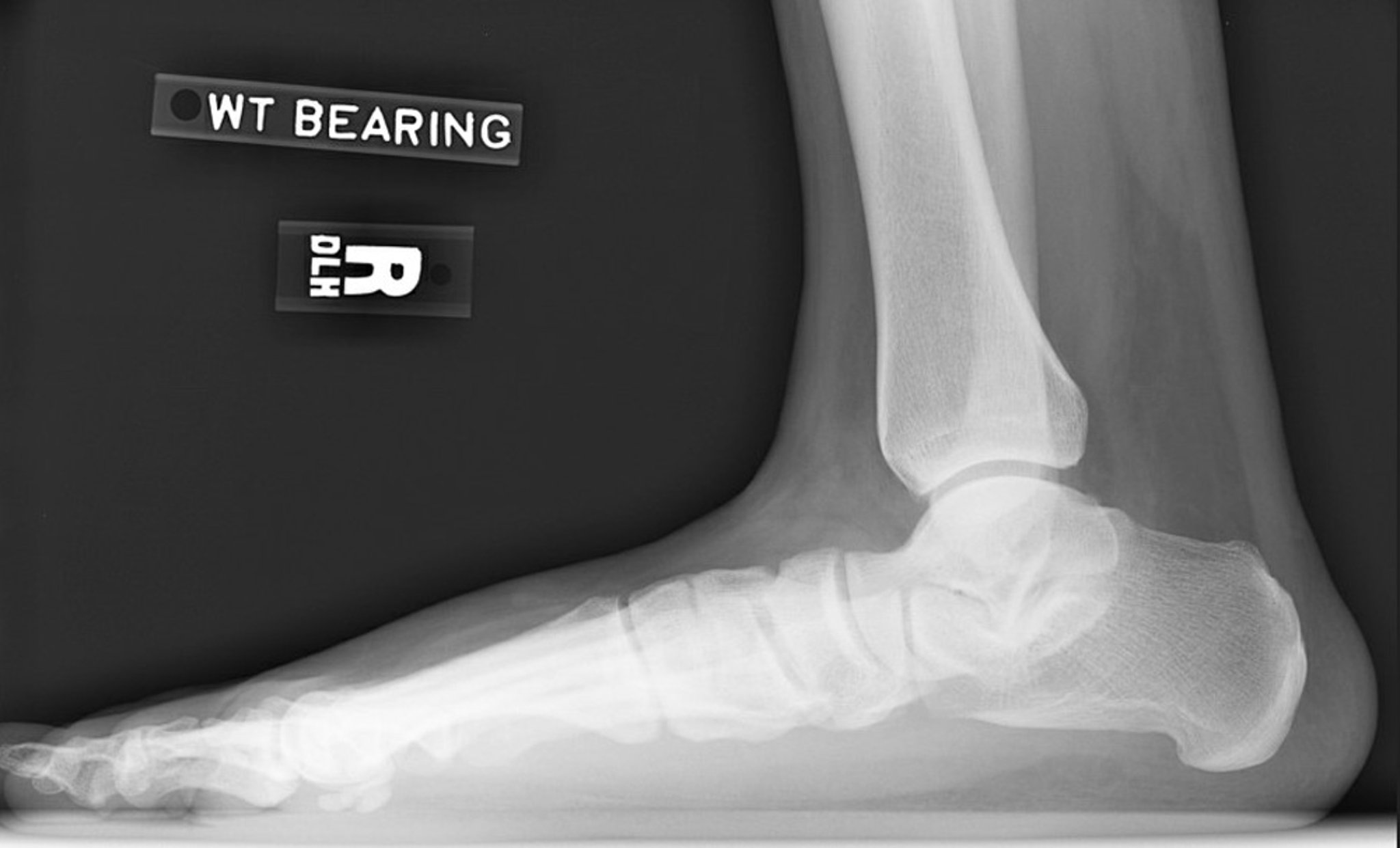
Lateral radiograph of right foot showing loss of arch height. Note the decreased calcaneal inclination angle and decreased talar declination angle. The subtalar joint is narrowed but the ankle joint is relatively preserved.
Image courtesy of James C. Connors, DPM.
Radiographs may be performed to exclude other structural abnormalities contributing to medial ankle pain (eg, os naviculare, an accessory bone that can become symptomatic). In addition, advanced tendinopathy can result in a collapsed foot arch, which on radiograph shows loss of arch height and joint malalignment of the subtalar, talonavicular, naviculocuneiform, and/or the calcaneocuboid joints. The calcaneus inclination angle is lost and the talar declination angle is flattened. The subtalar joint is narrowed due to calcaneal eversion.
MRI or ultrasound can confirm a fluid collection around the tendon (indicating tenosynovitis) or the extent of chronic degradation or tearing to the tendon with associated tendinosis.


Treatment
Orthotics and braces or surgery
Complete rupture of the tibialis posterior tendon requires surgery if normal function is the goal. Surgery is especially important in young active patients with acute tears.
Conservative therapy consists of mechanically off-loading the tendon by using orthotics modified with a deepened heel cup and appropriate medial wedging or posting in less severe pathology. Custom-molded ankle and foot bracing that extends the leg for added stability is indicated in more severe pathology.
Glucocorticoid injections exacerbate the degenerative process (see Considerations for Using glucocorticoid Injections).
For tenosynovitis, rest and aggressive anti-inflammatory therapy are warranted.


