

Stevens-Johnson syndrome and toxic epidermal necrolysis are severe cutaneous hypersensitivity reactions. Medications, especially sulfa drugs, antiseizure medications, and antibiotics, are the most common causes. Macules rapidly spread and coalesce, leading to epidermal blistering, necrosis, and sloughing. Diagnosis is usually obvious by appearance of initial lesions and clinical syndrome. Treatment is supportive care; cyclosporine, plasmapheresis or IV immune globulin, early corticosteroid therapy, and tumor necrosis factor-alpha inhibitors have been used. Stevens-Johnson syndrome and toxic epidermal necrolysis are severe cutaneous hypersensitivity reactions. Medications, especially sulfa drugs, antiseizure medications, and antibiotics, are the most common causes. Macules rapidly spread and coalesce, leading to epidermal blistering, necrosis, and sloughing. Diagnosis is usually obvious by appearance of initial lesions and clinical syndrome. Treatment is supportive care; cyclosporine, plasmapheresis or IV immune globulin, early corticosteroid therapy, and tumor necrosis factor-alpha inhibitors have been used.
Stevens-Johnson syndrome (SJS) and toxic epidermal necrolysis (TEN) are clinically similar except for their distribution. By one commonly accepted definition, changes affect < 10% of body surface area in SJS and > 30% of body surface area in TEN; involvement of 10 to 30% of body surface area is considered SJS/TEN overlap.
The disorders affect between 2 and 9 people/million (1). Incidence, severity, or both of these disorders may be higher in recipients of bone marrow transplants, in patients with HIV who have Pneumocystis jirovecii infection, in patients with systemic lupus erythematosus, and in patients with other chronic rheumatologic diseases.
General reference
1. Hsu DY, Brieva J, Silverberg NB, Silverberg JI. Morbidity and Mortality of Stevens-Johnson Syndrome and Toxic Epidermal Necrolysis in United States Adults. J Invest Dermatol. 2016;136(7):1387-1397. doi:10.1016/j.jid.2016.03.023
Etiology of SJS and TEN
Medications precipitate most cases of SJS and TEN. The most common medication causes include
Sulfa drugs (eg, sulfasalazine)
Other antibiotics (eg, aminopenicillins [usually ampicillin or amoxicillin], fluoroquinolones, cephalosporins)
Antiseizure medications (eg, phenytoin, carbamazepine, phenobarbital, valproic acid and its derivatives, lamotrigine)
Nonsteroidal anti-inflammatory drugs (eg, piroxicam, meloxicam)
Antiretroviral medications (eg, nevirapine)
Miscellaneous individual medications (eg, allopurinol, chlormezanone)
Immune checkpoint inhibitors (1)
Cases that are not caused by medications are attributed to
Infection (mostly with Mycoplasma pneumoniae)
Vaccination
In children with Stevens-Johnson syndrome, an infection is the most likely cause, and most of those are related to M. pneumoniae.
Rarely, a cause cannot be identified.
Etiology reference
1. Zhu J, Chen G, He Z, et al. Stevens-Johnson syndrome/toxic epidermal necrolysis in patients treated with immune checkpoint inhibitors: A safety analysis of clinical trials and FDA pharmacovigilance database. EClinicalMedicine. 2021;37:100951. Published 2021 Jun 10. doi:10.1016/j.eclinm.2021.100951
Pathophysiology of SJS and TEN
The exact mechanism of Stevens-Johnson syndrome and toxic epidermal necrolysis is unknown; however, one theory holds that altered medication metabolism (eg, failure to clear reactive metabolites) in some patients triggers a T-cell–mediated cytotoxic reaction to medication antigens in keratinocytes. CD8+ T cells have been identified as important mediators of blister formation. Findings suggest that granulysin released from cytotoxic T cells and natural killer cells might play a role in keratinocyte death; granulysin concentration in blister fluid correlates with severity of disease. Interleukin-15 has also been found to be increased in patients with SJS/TEN and has been found to increase granulysin production.
Another theory is that interactions between Fas (a cell-surface receptor that induces apoptosis) and its ligand, particularly a soluble form of Fas ligand released from mononuclear cells, lead to cell death and blister formation.
A genetic predisposition for SJS/TEN has been suggested.
Symptoms and Signs of SJS and TEN
Within 1 to 3 weeks after the start of the offending medication, patients develop a prodrome of malaise, fever, headache, cough, and keratoconjunctivitis. Macules, often in a target configuration, then appear suddenly, usually on the face, neck, and upper trunk. These macules simultaneously appear elsewhere on the body, coalesce into large flaccid bullae, and slough over a period of 1 to 3 days. Nails and eyebrows may be lost along with epithelium. The palms and soles may be involved. Skin, mucosal, and eye pain are common. In some cases, diffuse erythema is the first skin abnormality of TEN.

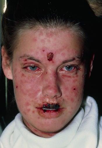
This photo shows an erythematous rash and blisters on the skin and on the mucosa of the eyes and mouth in this patient with SJS.


DR M.A. ANSARY/SCIENCE PHOTO LIBRARY
In severe cases of TEN, large sheets of epithelium slide off the entire body at pressure points (Nikolsky sign), exposing weepy, painful, and erythematous skin. Painful oral crusts and erosions, keratoconjunctivitis, and genital problems (eg, urethritis, phimosis, vaginal synechiae) accompany skin sloughing in most cases. Bronchial epithelium may also slough, causing cough, dyspnea, pneumonia, pulmonary edema, and hypoxemia. Glomerulonephritis and hepatitis may develop.
Diagnosis of SJS and TEN
Clinical evaluation
Often skin biopsy
Diagnosis is often obvious from appearance of lesions and rapid progression of symptoms. Histologic examination of sloughed skin shows necrotic epithelium, a distinguishing feature.
Differential diagnosis in Stevens-Johnson syndrome (SJS) and early toxic epidermal necrolysis (TEN) includes erythema multiforme, viral exanthems, and other drug rashes; SJS/TEN can usually be differentiated clinically as the disorder evolves and is characterized by significant pain and skin sloughing. In later stages of TEN, differential diagnosis includes the following:
Toxic shock syndrome (usually has more prominent multiorgan involvement and different cutaneous manifestations, such as macular rash on palms and soles that evolves to desquamation over about 2 weeks)
Exfoliative erythroderma (usually spares mucous membranes and is not as painful)
Paraneoplastic pemphigus (sometimes with different mucocutaneous findings or in patients with evidence of cancer)
In children, TEN is less common and must be distinguished from staphylococcal scalded skin syndrome. Characteristics of staphylococcal scalded skin syndrome usually include sparing of mucous membranes, absence of risk factors for TEN (eg, medication history), and clinical suspicion of staphylococcal infection.
Treatment of SJS and TEN
Supportive care
CyclosporineCyclosporine
Possibly corticosteroids, plasmapheresis, IV immune globulin (IVIG), or tumor necrosis factor (TNF)-alpha inhibitors
Treatment is most successful when Stevens-Johnson syndrome and toxic epidermal necrolysis (SJS/TEN) are recognized early and treated in an inpatient dermatologic or intensive care unit setting; treatment in a burn unit may be needed for severe disease (1).
Ophthalmology consultation and specialized eye care are mandatory for patients with ocular involvement. Potentially causative medications should be stopped immediately. Patients are isolated to minimize exposure to infection and are given fluids, electrolytes, blood products, and nutritional supplements as needed. Skin care includes prompt treatment of secondary bacterial infections and daily wound care as for severe burns. Prophylactic systemic antibiotics are controversial and are often avoided.
Pharmacotherapy of SJS/TEN is controversial. Cyclosporine (3 to 5 mg/kg orally once/day) inhibits CD8 cells and has been shown to decrease the duration of active disease (eg, by 2 to 3 days in some instances) and possibly decrease mortality (Pharmacotherapy of SJS/TEN is controversial. Cyclosporine (3 to 5 mg/kg orally once/day) inhibits CD8 cells and has been shown to decrease the duration of active disease (eg, by 2 to 3 days in some instances) and possibly decrease mortality (2). The use of systemic corticosteroids remains controversial. Many experts thought systemic corticosteroids increased mortality because of increased rates of infection and the risk of masking sepsis. However, some reports show improved outcomes with early corticosteroid therapy (3).
Plasmapheresis can remove reactive drug metabolites or antibodies and can be considered.
Early high-dose IVIG 2.7 g/kg over 3 days blocks antibodies and Fas ligand. However, despite some remarkable initial results using high-dose IVIG for TEN, further clinical trials involving small cohorts have reported conflicting results, and a retrospective analysis has suggested no improvement or even higher than expected mortality (4).
The TNF-alpha inhibitors infliximab and etanercept can help reduce inflammation.The TNF-alpha inhibitors infliximab and etanercept can help reduce inflammation.
Thalidomide has also been tested but increases mortality and is now contraindicated (5).
Treatment references
1. Seminario-Vidal L, Kroshinsky D, Malachowski SJ, et al. Society of Dermatology Hospitalists supportive care guidelines for the management of Stevens-Johnson syndrome/toxic epidermal necrolysis in adults. J Am Acad Dermatol. 2020;82(6):1553-1567. doi:10.1016/j.jaad.2020.02.066
2. Ng QX, De Deyn MLZQ, Venkatanarayanan N, Ho CYX, Yeo WS. A meta-analysis of cyclosporine treatment for Stevens-Johnson syndrome/toxic epidermal necrolysis. J Inflamm Res. 2018;11:135-142. Published 2018 Mar 28. doi:10.2147/JIR.S160964
3. Zimmermann S, Sekula P, Venhoff M, et al. Systemic Immunomodulating Therapies for Stevens-Johnson Syndrome and Toxic Epidermal Necrolysis: A Systematic Review and Meta-analysis. JAMA Dermatol. 2017;153(6):514-522. doi:10.1001/jamadermatol.2016.5668
4. Kirchhof MG, Miliszewski MA, Sikora S, et al. Retrospective review of Stevens-Johnson syndrome/toxic epidermal necrolysis treatment comparing intravenous immunoglobulin with cyclosporine. J Am Acad Dermatol. 2014;71(5):941–947. doi:10.1016/j.jaad.2014.07.016
5. Wolkenstein P, Latarjet J, Roujeau JC, et al. Randomised comparison of thalidomide versus placebo in toxic epidermal necrolysis. Lancet. 1998;352(9140):1586-1589. doi:10.1016/S0140-6736(98)02197-7
Prognosis for SJS and TEN
Severe toxic epidermal necrolysis is similar to extensive burns; patients are acutely ill, may be unable to eat or open their eyes, and suffer massive fluid and electrolyte losses. They are at high risk of infection, multiorgan failure, and death.
Mortality can be as high as 25 to 35% in adults but tends to be lower in children and with early treatment (1).
With early therapy, survival rates for TEN approach 90%. The (SCORTEN) systematically scores 7 independent risk factors within the first 24 hours of presentation to the hospital to determine the mortality rate for a particular patient.
Severity-of-Illness Score for Toxic Epidermal Necrolysis (SCORTEN)
Risk Factor* | Score | |
|---|---|---|
0 | 1 | |
Age | < 40 years | ≥ 40 years |
Associated cancer | No | Yes |
Heart rate (beats/minute) | < 120 | ≥ 120 |
Serum blood urea nitrogenSerum blood urea nitrogen | ≤ 28 mg/dL (10 mmol/L) | > 28 mg/dL (10 mmol/L) |
Detached or compromised body surface | < 10% | ≥ 10% |
Serum bicarbonate | ≥ 20 mEq/L (≥ 20 mmol/L) | < 20 mEq/L (< 20 mmol/L) |
Serum glucose | ≤ 250 mg/dL (≤ 13.88 mmol/L) | > 250 mg/dL (> 13.88 mmol/L) |
* More risk factors indicate a higher score and a higher mortality rate (%) as follows:
| ||
CI = confidence interval. | ||
Data from Bastuji-Garin S, Fouchard N, Bertocchi M, et al: SCORTEN: A severity-of-illness score for toxic epidermal necrolysis. J Invest Dermatol 115:149–153, 2000. doi: 10.1046/j.1523-1747.2000.00061.x | ||
Prognosis reference
1. Del Pozzo-Magaña BR, Lazo-Langner A. Stevens-Johnson Syndrome and Toxic Epidermal Necrolysis in Children: A Literature Review of Current Treatments. EMJ Dermatol. 2016;4[1]:83-89. doi:10.33590/emjdermatol/10314211
Key Points
Medications cause most cases of Stevens-Johnson syndrome (SJS) and toxic epidermal necrolysis (TEN), but infection, vaccination, and graft-vs-host disease are also potential causes.
Confirm the diagnosis by biopsy (showing necrotic epithelium) if clinical characteristics (eg, target lesions progressing to bullae, ocular and mucous membrane involvement, Nikolsky sign, desquamation in sheets) are inconclusive.
Early treatment decreases the often high mortality rate.
Except for mild cases, treat SJS/TEN in a burn unit and with intensive supportive care.
Consult ophthalmology if the eyes are affected.
Consider cyclosporine and possibly plasmapheresis for severe cases.Consider cyclosporine and possibly plasmapheresis for severe cases.


