


Postinfectious Glomerulonephritis (Immunofluorescent Staining)

Postinfectious Glomerulonephritis (Immunofluorescent Staining)
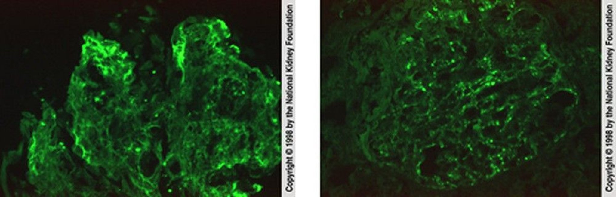
In the left image, immunofluorescent staining with anti-IgG demonstrates irregular IgG deposition in capillary loops (×400). In the right image, immunofluorescent staining with anti-C3 demonstrates scattered granular C3 deposition in capillary walls (×400).
Image provided by Agnes Fogo, MD, and the American Journal of Kidney Diseases' Atlas of Renal Pathology (see www.ajkd.org).
In these topics