


Risk factors for complications during pregnancy include
Preexisting maternal medical conditions and characteristics
Modifiable risk factors (eg, smoking, substance use)
Previous obstetric complications (eg, a previous history of preeclampsia)
Some major risk factors are discussed here. For additional medical conditions that may complicate pregnancy, see Pregnancy Complicated by Disease.
Diabetes
Preexisting diabetes mellitus is present in approximately 1% of pregnancies (1, 2), and gestational diabetes occurs in approximately 8% of pregnancies (3). Incidence is increasing as the incidence of obesity increases.
Preexisting insulin-dependent diabetes increases the risk of the following:
Fetal death
Major fetal malformations
Fetal macrosomia (fetal weight > 4.5 kg)
If vasculopathy is present, fetal growth restriction
The need for preterm, cesarean, or operative delivery
The incidence of fetal macrosomia is approximately 50% higher in pregnant women with preexisting diabetes than in pregnant women in the general population. The incidence of perinatal fetal or neonatal mortality is also higher.
Women with preexisting diabetes are more likely to require preterm delivery for obstetric or medical indications. Exercise during pregnancy (with judicious changes in diet) reduces the need for cesarean and operative deliveries in these women (4, 5).
Tight glucose control before conception and during early pregnancy is essential to prevent fetal malformations.
Insulin requirements usually increase during pregnancy.
Gestational diabetes increases the risk of the following:
Hypertensive disorders of pregnancy
The need for cesarean delivery
Gestational diabetes is routinely screened for at 24 to 28 weeks and, if women have risk factors, during the first trimester. Risk factors include the following:
Previous gestational diabetes
A macrosomic infant in a previous pregnancy
Unexplained fetal losses
Prepregnancy body mass index (BMI) > 30 kg/m2
Maternal age > 40 years
Family history of diabetes
Non-Hispanic Asian/Pacific Islander and Hispanic/Latina ethnicity
Screening and confirmation of the diagnosis of gestational diabetes can be done in 1 or 2 steps (6, 7):
1-step test: A fasting, 75-g glucose, 2-hour oral glucose tolerance test (GTT). Abnormal results are any of the following: fasting (≥ 92 mg/dL [5.1 mmol/L]); 1 hour (≥ 180 mg/dL [10 mmol/L]); or 2 hour (≥ 153 mg/dL [8.5 mmol/L).
2-step test: A non-fasting, 50-g glucose, 1-hour GTT; if abnormal (≥ 130 mg/dL [7.5 mmol/L] to 140 mg/dL [7.8 mmol/L]), then a fasting, 100-g, 3-hour GTT. For interpretation of results , see table Glucose Thresholds for Gestational Diabetes Mellitus Using a 3-hour Oral Glucose Tolerance Test.
The American College of Obstetricians and Gynecologists (ACOG) recommends the 2-step test to diagnosis gestational diabetes. The 1-step approach has been used and promoted by other organizations, including the International Association of Diabetes and Pregnancy Study Group (IADPSG) (6).
Optimal treatment of gestational diabetes (with dietary modification, exercise, and close monitoring of blood glucose levels and insulin when necessary) reduces risk of adverse maternal, fetal, and neonatal outcomes. Women with gestational diabetes are at a higher lifetime risk of cardiovascular events and, after delivery, should be referred for appropriate cardiovascular risk assessment and follow-up.
Women with gestational diabetes mellitus may have had undiagnosed diabetes mellitus before pregnancy. Thus, they should be screened for diabetes mellitus 6 to 12 weeks postpartum, using the same testing and criteria used for patients who are not pregnant.
See Diabetes Mellitus in Pregnancy for details regarding management of diabetes in pregnancy.
Diabetes references
1. Deputy NP, Kim SY, Conrey EJ, Bullard KM: Prevalence and Changes in Preexisting Diabetes and Gestational Diabetes Among Women Who Had a Live Birth - United States, 2012-2016. MMWR Morb Mortal Wkly Rep 67(43):1201-1207, 2018. Published 2018 Nov 2. doi:10.15585/mmwr.mm6743a2
2. Goya M, Codina M: Diabetes mellitus and pregnancy. Updated clinical practice guideline 2021. Executive summary. Endocrinol Diabetes Nutr (Engl Ed) 70 Suppl 1:1-6, 2023. doi:10.1016/j.endien.2021.12.006
3. Gregory EC, Ely DM: Trends and Characteristics in Gestational Diabetes: United States, 2016-2020. Natl Vital Stat Rep 71(3):1-15, 2022.
4. Artal R: Exercise: The alternative therapeutic intervention for gestational diabetes. Clinical Obstetrics and Gynecology 46 (2):479–487, 2003.
5. Artal R: The role of exercise in reducing the risks of gestational diabetes mellitus in obese women. Best Pract Res Clin Obstet Gynaecol 29 (1):123–4132, 2015.
6. American College of Obstetrics and Gynecology (ACOG): ACOG Practice Bulletin No. 190: Gestational Diabetes Mellitus. Obstet Gynecol. 2018;131(2):e49-e64. doi:10.1097/AOG.0000000000002501
7. American Diabetes Association Professional Practice Committee: 2. Diagnosis and Classification of Diabetes: Standards of Care in Diabetes-2024. Diabetes Care. 2024;47(Suppl 1):S20-S42. doi:10.2337/dc24-S002
Hypertension
Hypertensive disorders of pregnancy are classified as (1)
Chronic hypertension: Present before the pregnancy or developing before 20 weeks of pregnancy
Gestational hypertension: New onset of systolic and/or diastolic blood pressure (BP) ≥ 140/≥ 90 mm Hg on 2 occasions at least 4 hours apart after 20 weeks of gestation
Preeclampsia: New onset after 20 weeks of gestation of persistent (2 episodes within 4 hours) systolic and/or diastolic BP ≥ 140/≥ 90 mm Hg OR at least 1 measurement of systolic and/or diastolic BP ≥ 160/≥ 110 mm Hg PLUS new unexplained proteinuria (> 300 mg/24 hours or urine protein/creatinine ratio ≥ 0.3 or dipstick reading of 2+; in the absence of proteinuria, new-onset hypertension with new onset of other signs of end-organ damage (eg, thrombocytopenia [platelets < 100,000/mcL], impaired liver function, renal insufficiency, pulmonary edema, new-onset headache [unresponsive to medication and not accounted for by alternative diagnoses], visual symptoms).
Preeclampsia with severe features: Preeclampsia with persistent (2 episodes within 4 hours) systolic and/or diastolic BP ≥ 160/≥ 110 mm Hg and/or other signs of end-organ damage
HELLP syndrome: A form of severe preeclampsia with hemolysis, elevated liver enzymes, and low platelet count
Chronic hypertension plus superimposed preeclampsia: Worsening hypertension and new or worsening proteinuria or other signs of end-organ damage after 20 weeks in a woman with preexisting hypertension
Eclampsia: New-onset tonic-clonic, focal, or multifocal seizures not accounted for by other causes
Chronic hypertension increases risk of the following:
Fetal growth restriction (by decreasing uteroplacental blood flow)
Adverse fetal and maternal outcomes
Before attempting to become pregnant, women with hypertension should be counseled about risks. Prenatal care should begin as early in pregnancy as possible. Pregnant women with chronic hypertension who require antihypertensive therapy should be started or continued on appropriate medications and referred to a maternal-fetal medicine specialist (2).
Management of chronic hypertension during pregnancy
Ultrasonography to monitor fetal growth is done at 28 weeks and every 4 weeks thereafter. Delayed growth is evaluated with multivessel Doppler testing by a maternal-fetal medicine specialist.
Women with a history of preeclampsia or gestational hypertension are at a higher lifetime risk of cardiovascular events and, after delivery, should be referred for appropriate cardiovascular risk assessment and follow-up.
Hypertension references
1. American College of Obstetrics and Gynecology (ACOG): ACOG Practice Bulletin, Number 222: Gestational hypertension and preeclampsia. Obstet Gynecol 133 (1):1, 2019. doi: 10.1097/AOG.0000000000003018
2. Tita AT, Szychowski JM, Boggess K, et al: Treatment for Mild Chronic Hypertension during Pregnancy. N Engl J Med 386(19):1781-1792, 2022. doi:10.1056/NEJMoa2201295
3. American College of Obstetrics and Gynecology (ACOG)Obstet Gynecol 132 (1):e44–e52, 2018. doi: 10.1097/AOG.0000000000002708
4. Ayyash M, Goyert G, Garcia R, et alAm J Perinatol. Published online July 29, 2023. doi:10.1055/s-0043-1771260
Thyroid Disorders
Thyroid disorders may predate or develop during pregnancy. Pregnancy does not change the symptoms of hypothyroidism and hyperthyroidism.
Fetal effects vary with the disorder and the medications used for treatment. But generally, untreated or inadequately treated hyperthyroidism can result in
Untreated hypothyroidism can cause
Intellectual deficits in children
The most common causes of maternal hypothyroidism are Hashimoto thyroiditis and treatment of Graves disease.
If women have or have had a thyroid disorder, thyroid status should be closely monitored during and after pregnancy in the women and after delivery in infants. Goiters and thyroid nodules discovered during pregnancy should be evaluated as they are in nonpregnant patients (see Approach to the Patient With a Thyroid Nodule and Diagnosis of Simple Nontoxic Goiter).
Sexually Transmitted Infections (STIs)
(See also Sexually Transmitted Infections and Infectious Disease in Pregnancy.)
Screening for sexually transmitted infections should be done during pregnancy to make treatment possible and to prevent adverse effects of intrauterine or perinatal transmitted infections to the fetus or neonate. The rate of congenital syphilis in the United States consistently rises (see CDC: National Overview of STDs, 2021).
Routine prenatal care includes screening tests for HIV infection, hepatitis B, hepatitis C (1), and syphilis and, if < 25 years, for chlamydial infection and gonorrhea at the first prenatal visit. Syphilis testing is repeated during pregnancy and at delivery if risk continues or if the patient resides in an endemic area (2). Pregnant women who have any of these infections are treated with antimicrobials.
Fetal syphilis in utero can cause fetal death, congenital malformations, and severe disability.
Without treatment, risk of transmission of HIV from mother to child is approximately 30% prepartum and approximately 25% intrapartum. Antiretroviral treatment of the pregnant woman before and during pregnancy and of the neonate within 6 to 12 hours of birth reduces risk of HIV transmission to the fetus by two thirds; risk is lower (< 2%) with a combination of 2 or 3 highly active antiretrovirals. Highly active antiretrovirals are given to the mother during pregnancy and intrapartum to prevent mother-to-child transmission.
During pregnancy, hepatitis, bacterial vaginosis, gonorrhea, and genital chlamydial infection increase risk of preterm labor and prelabor rupture of the membranes.
Treatment of bacterial vaginosis, gonorrhea, or chlamydial infection may prolong the interval from rupture of the membranes to delivery and may improve fetal outcome by decreasing fetal inflammation.
STIs references
1. American College of Obstetrics and Gynecology (ACOG): ACOG Clinical Practice Guideline No. 6.: Viral Hepatitis in Pregnancy, Obstet Gynecol 2023;142(3):745-759. doi:10.1097/AOG.0000000000005300
2. Workowski KA. , Laura H. Bachmann LH, Chan PA: Sexually transmitted infections treatment guidelines, 2021. MMWR Recomm Rep 70 (4):1–187, 2021. doi: http://dx.doi.org/10.15585/mmwr.rr7004a1external icon
Female Genital Tract Abnormalities
Structural abnormalities of the uterus and cervix (eg, uterine septum, bicornuate uterus) make the following more likely:
Spontaneous abortion during the second trimester
Preterm labor or delivery
Dysfunctional labor
The need for cesarean delivery
Uterine fibroids uncommonly cause placental abnormalities (eg, placenta previa), preterm labor, and recurrent spontaneous abortion. Fibroids may grow rapidly or degenerate during pregnancy; degeneration often causes severe pain and peritoneal signs and may also cause preterm labor.
Cervical insufficiency (incompetence) makes preterm delivery more likely. The risk of cervical insufficiency is higher in women who have had lacerations or injury of the cervix during a previous procedure (eg, therapeutic abortion, instrumental vaginal deliveries). Cervical insufficiency is treated with surgical intervention (cerclage).
If, before pregnancy, women have had a myomectomy in which the uterine cavity was entered, cesarean delivery is required because uterine rupture is a risk during subsequent vaginal delivery.
Uterine abnormalities that lead to poor obstetric outcomes often require surgical correction, which is done after delivery.
Substance Use
Substance use during pregnancypreterm labor, low birth weight, congenital anomalies, developmental delays, and long-term behavioral and cognitive problems in the child.
Maternal substance use, especially with opioids, can also result in neonatal withdrawal symptoms requiring specialized care.
Alcohol is the most commonly used teratogen. Risk is probably related to amount of alcohol consumed, but no amount is known to be risk-free. Binge drinking in particular, possibly as little as 45 mL of pure alcohol (equivalent to about 3 drinks) a day, can cause fetal alcohol syndrome, which may result in fetal growth restriction, facial and cardiovascular defects, and neurologic dysfunction.
Clinicians should ask pregnant patients about substance misuse, utilize validated screening tools, and refer to addiction specialists when necessary.
Exposure to Teratogens
Common teratogens (agents that cause fetal malformation) include infections, drugs, and physical agents. Malformations are most likely to result if exposure occurs between the second and eighth week after conception (the fourth to tenth week after the last menstrual period), when organs are forming. Other adverse pregnancy outcomes are also more likely. Pregnant women exposed to teratogens are counseled about increased risks and referred for detailed ultrasound evaluation to detect malformations.
Common infections that may be teratogenic include
Commonly used drugs that may be teratogenic include
Bath salts (cathinones)
Some prescription drugs (see table Drugs With Adverse Effects During Pregnancy)
Hyperthermia or exposure to temperatures > 39° C (eg, in a sauna) during the first trimester has been associated with spina bifida.
Exposure to Mercury
Mercury in seafood can be toxic to the fetus. The FDA (see Advice about Eating Fish For Those Who Might Become or Are Pregnant or Breastfeeding and Children Ages 1–11 Years) recommends the following:
Avoiding tilefish from the Gulf of Mexico, shark, swordfish, big-eye tuna, marlin, orange roughy, and king mackerel
Limiting albacore tuna to 4 ounces (one average meal)/week
Before eating fish caught in local lakes, rivers, and coastal areas, checking local advisories about the safety of such fish and, if levels of mercury are not known to be low, limiting consumption to 4 ounces/week while avoiding other seafood that week
Tilefish from the Gulf of Mexico have the highest levels of mercury of all fish (as tested by the U.S. Food and Drug Administration (FDA); tilefish from the Atlantic Ocean can be safely eaten.

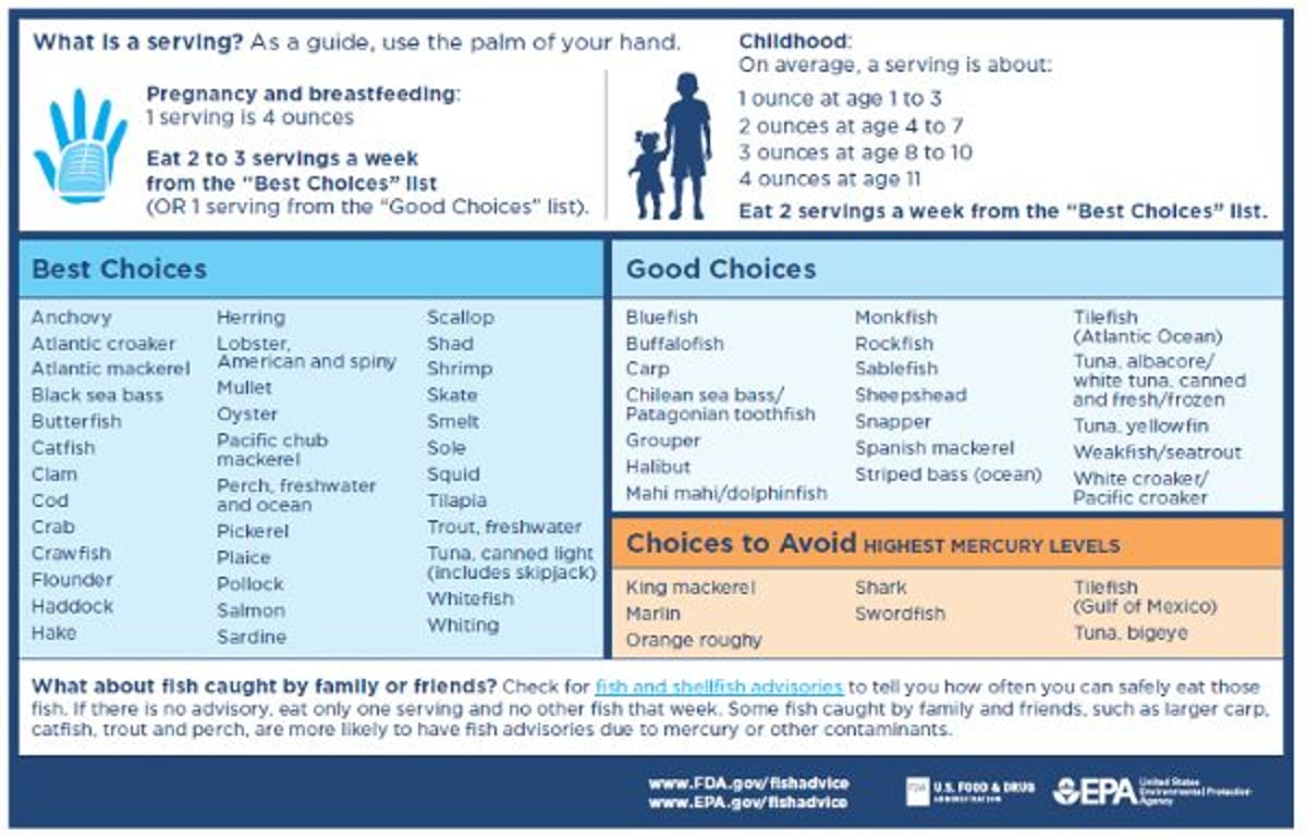
Image from the U.S. Food and Drug Administration (FDA) and the United States Environmental Protection Agency (EPA).
Experts recommend that women who are pregnant or breastfeeding eat 8 to 12 ounces (2 or 3 average meals) per week of a variety of seafood that is lower in mercury. Such seafood includes flounder, shrimp, canned light tuna, salmon, pollock, tilapia, cod, and catfish. Fish has nutrients that are important for fetal growth and development.
Maternal Age
Adolescents, who account for 13% of all pregnancies, have an increased incidence of preeclampsia, preterm labor, and anemia, which often leads to fetal growth restriction. The cause, at least in part, is that adolescents tend to neglect prenatal care, frequently smoke cigarettes or use other substances, and have higher rates of sexually transmitted infections.
In women ≥ 35 years, the incidence of preeclampsia is increased, as is that of gestational diabetes, dysfunctional labor, abruptio placentae, stillbirth, and placenta previa. These women are also more likely to have preexisting disorders (eg, chronic hypertension, diabetes). Because risk of fetal chromosomal abnormalities increases as maternal age increases, genetic screening and testing and detailed ultrasound screening for fetal malformations should be offered.
The most common chromosomal abnormality is autosomal trisomy. The United States National Birth Defects Prevention Study (NBDPS) found that children of women > 40 years are at increased risk of cardiac abnormalities, esophageal atresia, hypospadias, and craniosynostosis (1).
Maternal age reference
1. Gill SK, Broussard C, Devine O, et al: Association between maternal age and birth defects of unknown etiology: United States, 1997-2007. Birth Defects Res A Clin Mol Teratol 94 (12):1010–1018, 2012. doi: 10.1002/bdra.23049
Maternal Weight
Pregnant women whose body mass index (BMI) was < 18.5 kg/m2 before pregnancy are considered underweight, which predisposes to low birth weight (< 2.5 kg) in neonates. Such women are encouraged to gain at least 12.5 kg during pregnancy.
Pregnant women whose BMI was 25 to 29.9 kg/m2 (overweight) or ≥ 30 kg/m2 (obese) before pregnancy are at risk of maternal hypertension and diabetes, postterm pregnancy, pregnancy loss, fetal macrosomia, congenital malformations, intrauterine growth restriction, preeclampsia, and the need for cesarean delivery. Ideally, weight loss should begin before pregnancy, first by trying lifestyle modifications (eg, increased physical activity, dietary changes). Women with overweight or obesity are encouraged to limit weight gain during pregnancy, ideally by modifying their lifestyle. The Institute of Medicine (IOM) uses the following guidelines:
Overweight: Weight gain limited to 6.8 to 11.3 kg (15 to 25 lb)
Obese: Weight gain limited to < 5 to 9 kg (11 to 20 lb)
However, not all experts agree with IOM recommendations. Many experts recommend an individualized approach that can include more limited weight gain plus lifestyle modifications (eg, increased physical activity, dietary changes), particularly for women with obesity (1). During pregnancy, most women should be encouraged to exercise at least 3 times a week for a total of 150 minutes each week (2).
For pregnant women with overweight or obesity, lifestyle modifications during pregnancy reduce the risk of gestational diabetes and preeclampsia.
Discussing appropriate weight gain, diet, and exercise at the initial visit and periodically throughout the pregnancy is important.
Maternal weight references
1. Artal R, Lockwood CJ, Brown HL: Weight gain recommendations in pregnancy and the obesity epidemic. Obstet Gynecol 115 (1):152–155, 2010. doi: 10.1097/AOG.0b013e3181c51908
2. Mottola MF, Davenport MH, Ruchat SM, et al: 2019 Canadian guideline for physical activity throughout pregnancy. Br J Sports Med 52 (21):1339–1346, 2018. doi: 10.1136/bjsports-2018-100056
Multiple Gestation
Multiple gestation increases risk of the following:
Congenital malformations
Perinatal morbidity and mortality
After delivery, uterine atony and hemorrhage
Multiple gestation is usually detected with ultrasonography in the first trimester. Incidence of multiple gestations has been increasing; use of assisted reproductive technologies have contributed substantially to this increase (1).
Multiple gestation reference
1. American College of Obstetricians and Gynecologists (ACOG): ACOG Practice Bulletin No. 231: Multifetal gestations: Twin, triplet, and higher-order multifetal pregnancies. Obstet Gynecol 137 (6):e145–e162, 2021. doi: 10.1097/AOG.0000000000004397
Prior Neonate With a Genetic or Congenital Disorder
Risk of having a fetus with a chromosomal disorder is increased for most couples who have had a fetus or neonate with a chromosomal disorder (recognized or missed). Recurrence risk for most genetic disorders is unknown. Most congenital malformations are multifactorial; risk of having a subsequent fetus with malformations varies based on the defect itself.
If couples have had a neonate with a genetic or chromosomal disorder, genetic consultation and screening is recommended. If couples have had a neonate with a congenital malformation, genetic screening, high-resolution ultrasonography, and evaluation by a maternal-fetal medicine specialist is recommended.
Prior Stillbirth
Stillbirth is death of a fetus at ≥ 20 weeks gestation before or during delivery, as defined by the Centers for Disease Control and Prevention (CDC [1]), or at > 28 weeks, as defined by the World Health Organization (2). Fetal death during late pregnancy may have maternal, placental, or fetal anatomic or genetic causes (see table Common Causes of Stillbirth). Having had a stillbirth or late abortion (ie, at 16 to 20 weeks) increases risk of fetal death in subsequent pregnancies. Degree of risk varies depending on the cause of a previous stillbirth. Fetal surveillance using antepartum testing (eg, nonstress testing, biophysical profile) is recommended.
Treatment of maternal disorders (eg, chronic hypertension, diabetes, infections) may lower risk of stillbirth in a current pregnancy.
Prior stillbirth references
1. CDC: What is stillbirth? Accessed March 2024.
2. World Health Organization: Stillbirth. Accessed March 2024.
Prior Preterm Delivery
Preterm delivery is delivery before 37 weeks. Previous preterm delivery due to preterm labor is associated with an increased risk of future preterm deliveries. Preterm delivery is also sometimes medically indicated due to certain pregnancy complications (eg, severe preeclampsia or symptomatic placenta previa). These diseases are not independent risk factors for preterm delivery, but they may recur, and appropriate monitoring or preventive measures should be done.
Women with prior preterm delivery due to unexplained preterm labor should be closely monitored at 2-week intervals beginning at 15 to 16 weeks gestation, up to 23 to 24 weeks. Evaluation may include (1)
Ultrasound evaluation, including measurement of cervical length, beginning at 15 to 16 weeks
Testing for bacterial vaginosis, if there are symptoms
Measurement of fetal fibronectin for women with symptoms concerning for preterm labor
Prior preterm delivery reference
1. Prediction and Prevention of Spontaneous Preterm Birth: ACOG Practice Bulletin, Number 234. Obstet Gynecol. 2021;138(2):e65-e90. doi:10.1097/AOG.0000000000004479
Prior Birth Injury
Most cerebral palsy and neurodevelopmental disorders are caused by factors unrelated to a birth injury. Injuries such as brachial plexus damage can result from procedures such as forceps or vacuum extractor delivery but often result from intrauterine forces during labor or malposition during the last weeks of pregnancy.
Previous shoulder dystocia is a risk factor for future dystocia, and the delivery records should be reviewed for potentially modifiable risk factors (eg, fetal macrosomia, operative vaginal delivery) that may have predisposed to the injury.


