

Chronic venous insufficiency is impaired venous return, sometimes causing lower extremity discomfort, edema, and skin changes. Post-thrombotic (postphlebitic) syndrome is symptomatic chronic venous insufficiency after deep venous thrombosis (DVT). Causes of chronic venous insufficiency are disorders that result in venous hypertension, usually as a result of venous damage or incompetence of venous valves, as occurs (for example) after DVT. Diagnosis is by history, physical examination, and duplex ultrasound. Treatment is compression, wound care, and, rarely, surgery. Prevention requires adequate treatment of DVT and the use of compression stockings.
Prevalence estimates of chronic venous insufficiency vary widely, reflecting differences in the study populations (1). Post-thrombotic syndrome may affect up to 50% of patients with deep venous thrombosis (DVT), and can have a substantial effect on quality of life. Post-thrombotic syndrome can range in severity from mild leg swelling to debilitating venous leg ulcers; approximately 5 to 23% of patients will develop moderate to severe disease. Post-thrombotic syndrome is more common in patients with more proximal and more extensive DVT, such as those with involvement of the common femoral and/or iliofemoral veins (2).
General references
1. Galanaud JP, Monreal M, Kahn SR. Epidemiology of the post-thrombotic syndrome. Thromb Res. 2018;164:100-109. doi:10.1016/j.thromres.2017.07.026
2. Rabinovich A, Kahn SR. How I treat the postthrombotic syndrome. Blood. 2018;131(20):2215-2222. doi:10.1182/blood-2018-01-785956
Etiology of Chronic Venous Insufficiency
Venous return from the lower extremities relies on contraction of the calf muscles to push blood from intramuscular (soleal) sinusoids and gastrocnemius veins into and through deep veins. Venous valves direct blood proximally to the heart. Chronic venous insufficiency occurs when venous obstruction (eg, in DVT), venous valvular insufficiency, lymphatic dysfunction, or decreased contraction of muscles surrounding the veins (eg, due to immobility) decrease forward venous flow and increase venous pressure (venous hypertension).
Fluid accumulation in the lower extremities (eg, in right heart failure) can also contribute by causing venous hypertension. Prolonged venous hypertension causes tissue edema, inflammation, and hypoxia, leading to symptoms. Pressure may be transmitted to superficial veins if valves in perforator veins, which connect deep and superficial veins, are ineffective.
Common risk factors for chronic venous insufficiency include (1):
Older age
Trauma
Occupations requiring prolonged standing or carrying heavy loads
Pregnancy
Idiopathic cases are often attributed to a history of occult DVT.
Post-thrombotic syndrome is symptomatic chronic venous insufficiency that follows DVT. Risk factors for post-thrombotic syndrome in patients with DVT include proximal thrombosis, recurrent ipsilateral DVT, and body mass index (BMI) ≥ 22 kg/m2. Age, female sex, and estrogen therapy are also associated with the syndrome.
Etiology reference
1. Fukaya E, Kolluri R. Nonsurgical Management of Chronic Venous Insufficiency. N Engl J Med. 2024;391(24):2350-2359. doi:10.1056/NEJMcp2310224
Symptoms and Signs of Chronic Venous Insufficiency
Clinically evident chronic venous insufficiency may not cause any symptoms but always causes signs; post-thrombotic syndrome always causes symptoms. Both disorders are a concern because their symptoms can mimic those of acute DVT and both can lead to substantial reductions in physical activity and quality of life.
Symptoms include a sense of fullness, heaviness, aching, cramps, pain, tiredness, and paresthesias in the legs; these symptoms worsen with standing or walking and are relieved by rest and elevation. Pruritus may accompany skin changes. Signs occur along a continuum: no changes to varicose veins (rare) to edema to stasis dermatitis on the lower legs and at the ankles, with or without ulceration (see table ). The calf may be painful when compressed.
Clinical Classification of Chronic Venous Insufficiency
Class | Signs |
|---|---|
C0 | No clinical signs of venous disease |
C1 | Telangiectatic or reticular veins* |
C2 | Varicose veins* C2r: Recurrent varicose veins |
C3 | Edema |
C4 | Skin changes due to venous stasis C4a: Pigmentation or eczema C4b: Lipodermatosclerosis† or atrophie blanche‡ C4c: Corona phlebectatica§ |
C5 | Healed venous ulcer |
C6 | Active venous ulcer C6r: Recurrent active venous ulcer |
* May occur idiopathically without chronic venous insufficiency. | |
† Inflammation with induration, discoloration, and edema | |
‡ Small, white depressed scars | |
§ Fan-shaped pattern of small intradermal veins on the medial or lateral ankle and foot | |
Data from Lurie F, Passman M, Meisner M, et al. The 2020 update of the CEAP classification system and reporting standards. J Vasc Surg Venous Lymphat Disord. 2020;8(3):342-352. doi:10.1016/j.jvsv.2019.12.075 | |
Venous stasis dermatitis consists of erythema, hyperpigmentation, induration, venous ectasia, lipodermatosclerosis (fibrosing subcutaneous panniculitis), lichenification, and venous stasis ulcers. Erythema may be difficult to appreciate in dark skin.

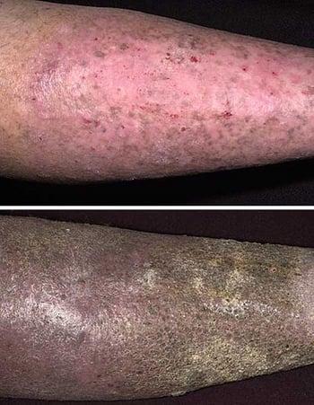
Chronic stasis dermatitis may appear as fibrotic skin thickening and hyperpigmentation. The changes are characteristic in both lightly pigmented skin (top) and darkly pigmented skin (bottom), here appearing more pronounced in the bottom photo.


BELMONTE/VABRES/SCIENCE PHOTO LIBRARY
Venous stasis ulcers may develop spontaneously or after affected skin is scratched or injured. They typically occur around the medial malleolus, tend to be shallow and moist, and may be malodorous (especially when poorly cared for) or painful. They do not penetrate the deep fascia. In contrast, ulcers due to peripheral artery disease eventually expose tendons or bone.

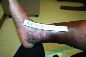
Venous stasis includes lichenification and hyperpigmentation. A shallow ulcer is developing superior to the medial malleolus.
Venous stasis includes lichenification and hyperpigmentation. A shallow ulcer is developing superior to the medial mall
© Springer Science+Business Media


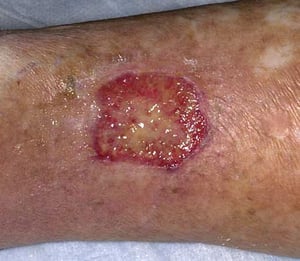
This large venous stasis ulcer is surrounded by brawny induration.
This large venous stasis ulcer is surrounded by brawny induration.
© Springer Science+Business Media

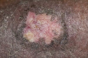
Venous stasis ulcers develop as a result of inadequately treated stasis dermatitis; they may quickly follow the first signs of stasis dermatitis.
Venous stasis ulcers develop as a result of inadequately treated stasis dermatitis; they may quickly follow the first s
Image provided by Thomas Habif, MD.
Roberto A. Penne-Casanova/SCIENCE PHOTO LIBRARY
Venous stasis includes lichenification and hyperpigmentation. A shallow ulcer is developing superior to the medial malleolus.
Venous stasis includes lichenification and hyperpigmentation. A shallow ulcer is developing superior to the medial mall
© Springer Science+Business Media
This large venous stasis ulcer is surrounded by brawny induration.
This large venous stasis ulcer is surrounded by brawny induration.
© Springer Science+Business Media
Venous stasis ulcers develop as a result of inadequately treated stasis dermatitis; they may quickly follow the first signs of stasis dermatitis.
Venous stasis ulcers develop as a result of inadequately treated stasis dermatitis; they may quickly follow the first s
Image provided by Thomas Habif, MD.
Roberto A. Penne-Casanova/SCIENCE PHOTO LIBRARY
Leg edema tends to be unilateral or asymmetric; bilateral symmetric edema is more likely to result from a systemic disorder (eg, heart failure, hypoalbuminemia) or certain medications (eg, calcium channel blockers).
In general, unless the lower extremities are adequately cared for, patients with any manifestation of chronic venous insufficiency or post-thrombotic syndrome are at risk of progression to more advanced disease.
Diagnosis of Chronic Venous Insufficiency
History and Physical Examination
Ultrasound to exclude DVT
Diagnosis is usually based on history and physical examination. The Villalta score, a clinical scoring system that ranks 5 symptoms (pain, cramps, heaviness, pruritus, paresthesia) and 6 signs (edema, hyperpigmentation, induration, venous ectasia, blanching hyperemia, pain with calf compression) on a scale of 0 (absent or minimal) to 3 (severe) is an effective diagnostic tool of disease severity (1, 2, 3, 4). Scores of 5 to 14 on 2 visits separated by ≥ 6 months indicate mild-to-moderate disease, and scores of ≥ 15 indicate severe disease.
Lower-extremity duplex ultrasound reliably excludes or confirms DVT. Absence of edema and a reduced ankle-brachial index suggest peripheral artery disease rather than chronic venous insufficiency or post-thrombotic syndrome.
Diagnosis references
1. Kahn SR. Measurement properties of the Villalta scale to define and classify the severity of the post-thrombotic syndrome. J Thromb Haemost. 2009;7(5):884-888. doi:10.1111/j.1538-7836.2009.03339.x
2. Kahn SR, Comerota AJ, Cushman M, et al. The postthrombotic syndrome: evidence-based prevention, diagnosis, and treatment strategies: a scientific statement from the American Heart Association. Circulation. 2014;130(18):1636-1661. doi:10.1161/CIR.0000000000000130
3. Ng S, Rodger MA, Ghanima W, et al. External Validation of the Patient-Reported Villalta Scale for the Diagnosis of Postthrombotic Syndrome. Thromb Haemost. 2022;122(8):1379-1383. doi:10.1055/a-1738-1313
4. O'Donnell TF Jr, Passman MA, Marston WA, et al. Management of venous leg ulcers: clinical practice guidelines of the Society for Vascular Surgery ® and the American Venous Forum. J Vasc Surg. 2014;60(2 Suppl):3S-59S. doi:10.1016/j.jvs.2014.04.049
Treatment of Chronic Venous Insufficiency
Elevation
Compression using bandages, stockings, and/or pneumatic devices
Exercise training
Topical treatments
Treatment of secondary infection, when present
Sometimes surgical and endovascular approaches
Weight loss, regular exercise, and reduction of dietary sodium may benefit patients with bilateral chronic venous insufficiency (1). Weight loss can also improve venous hypertension by reducing concomitant sleep apnea and right heart failure. Leg-strengthening exercises and aerobic exercise, preferably in a supervised program, may also benefit patients with post-thrombotic syndrome (2).
Elevating the leg above the level of the right atrium decreases venous hypertension and edema, is appropriate for all patients, and should be done a minimum of 3 times a day for ≥ 30 minutes. However, many patients cannot reliably adhere to this schedule during the day.
Compression is recommended for treatment and prevention of the effects of chronic venous insufficiency (ie, edema, venous ulcers) and can be used for all patients (1, 2). Although the data are mixed as to whether compression stockings prevent post-thrombotic syndrome, they are useful to reduce symptoms of swelling, pain, and tightness that may occur after deep vein thrombosis (2).
Elastic bandages are used initially until edema and ulcers resolve and leg size stabilizes; commercial compression stockings are then used. Stockings that provide 20 to 30 mm Hg of distal circumferential pressure are indicated for smaller varicose veins and mild chronic venous insufficiency; 30 to 40 mm Hg is indicated for larger varicose veins and moderate to severe disease. Infrequently, higher compression pressures (eg, > 40 mm Hg) can be used but may not be tolerated for long-term use. Stockings should be put on when patients awaken, before leg edema worsens with activity, and should exert maximal pressure at the ankles and gradually less pressure proximally. Adherence to this treatment varies; many patients consider stockings irritating, restricting, or cosmetically undesirable, and many patients have difficulty putting them on.
Intermittent pneumatic compression (IPC) uses a pump to cyclically inflate and deflate hollow plastic leggings. IPC provides external compression, squeezing blood and fluid out of the lower legs. It effectively treats severe post-thrombotic syndrome (2) and venous stasis ulcers but is less practical than compression stockings for patients to adhere to on an ongoing basis.
Options for venous ulcer management include compression, oral pentoxifylline, and topical treatments (include compression, oral pentoxifylline, and topical treatments (2). There is no strong evidence that one approach is superior, and management should be individualized. Involvement of a wound care specialist may be helpful to guide management. An Unna boot (zinc oxide–impregnated bandages) properly applied, covered by compression bandages, and changed weekly is associated with ulcer healing in some patients (). There is no strong evidence that one approach is superior, and management should be individualized. Involvement of a wound care specialist may be helpful to guide management. An Unna boot (zinc oxide–impregnated bandages) properly applied, covered by compression bandages, and changed weekly is associated with ulcer healing in some patients (3, 4). Occlusive dressings (eg, hydrocolloids such as aluminum chloride) provide a moist environment for wound healing and promote growth of new tissue; they may be used for ulcers with light to moderate exudate, but they probably add little to simple Unna bandaging. Dry dressings are absorptive, making them most appropriate for heavier exudate.). Occlusive dressings (eg, hydrocolloids such as aluminum chloride) provide a moist environment for wound healing and promote growth of new tissue; they may be used for ulcers with light to moderate exudate, but they probably add little to simple Unna bandaging. Dry dressings are absorptive, making them most appropriate for heavier exudate.
Other medications have little to no role in routine treatment of chronic venous insufficiency (2), although many patients are given aspirin, topical glucocorticoids, or antibiotics. Diuretics should be reserved for treating volume overload rather than local edema. If used, thiazides and mineralocorticoid receptor antagonists are preferred over loop diuretics because they can increase venous compliance (1).
Surgery (eg, venous ligation, stripping, valve reconstruction) has limited effectiveness (1). Grafting autologous skin or skin created from epidermal keratinocytes or dermal fibroblasts may be an option for patients with stasis ulcers refractory to all other measures (5); however, there is a risk that the graft may reulcerate, especially if there is ongoing venous hypertension. For patients with refractory venous ulcers, venous valve reconstruction may be considered (2). Surgical venovenous bypass procedures as well as endovascular stent venoplasty are options for patients with severe symptoms.
Treatment references
1. Fukaya E, Kolluri R. Nonsurgical Management of Chronic Venous Insufficiency. N Engl J Med. 2024;391(24):2350-2359. doi:10.1056/NEJMcp2310224
2. Kahn SR, Comerota AJ, Cushman M, et al. The postthrombotic syndrome: evidence-based prevention, diagnosis, and treatment strategies: a scientific statement from the American Heart Association [published correction appears in Circulation. 2015 Feb 24;131(8):e359]. Circulation. 2014;130(18):1636-1661. doi:10.1161/CIR.0000000000000130
3. Norman G, Westby MJ, Rithalia AD, Stubbs N, Soares MO, Dumville JC. Dressings and topical agents for treating venous leg ulcers. Cochrane Database Syst Rev. 2018;6(6):CD012583. doi:10.1002/14651858.CD012583.pub2
4. Paranhos T, Paiva CSB, Cardoso FCI, et al. Systematic review and meta-analysis of the efficacy of Unna boot in the treatment of venous leg ulcers. Wound Repair Regen. 2021;29(3):443-451. doi:10.1111/wrr.12903
5. Jones JE, Nelson EA, Al-Hity A. Skin grafting for venous leg ulcers. Cochrane Database Syst Rev. 2013;2013(1):CD001737. doi:10.1002/14651858.CD001737.pub4
Prevention of Chronic Venous Insufficiency
Primary prevention of chronic venous insufficiency involves DVT prophylaxis for those at risk, adequate anticoagulation after DVT and use of compression stockings for up to 2 years after DVT or lower extremity venous trauma. However, a meta-analysis of randomized trials comparing compression stockings with placebo (ie, either none or sham-compression stockings) failed to show a significant reduction in post-thrombotic syndrome (1).
Lifestyle changes (eg, weight loss, regular exercise) can decrease risk of chronic venous insufficiency by decreasing lower extremity venous pressure (2).
Prevention references
1. Subbiah R, Aggarwal V, Zhao H, Kolluri R, Chatterjee S, Bashir R. Effect of compression stockings on post thrombotic syndrome in patients with deep vein thrombosis: a meta-analysis of randomised controlled trials. Lancet Haematol. 2016;3(6):e293-e300. doi:10.1016/S2352-3026(16)30017-5
2. Fukaya E, Kolluri R. Nonsurgical Management of Chronic Venous Insufficiency. N Engl J Med. 2024;391(24):2350-2359. doi:10.1056/NEJMcp2310224
Key Points
Skin changes range on a continuum from normal skin or mildly ectatic veins to severe stasis dermatitis and ulceration.
Symptoms are more common with post-thrombotic syndrome and include heaviness, aching, and paresthesias.
Diagnosis is based on inspection, but patients should have ultrasound to exclude deep venous thrombosis.
Treatment is with elevation and compression; medications and surgery are typically ineffective.


