

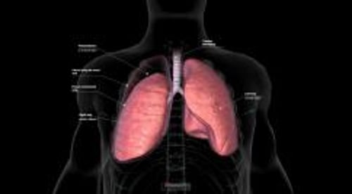
A pneumothorax is partial or complete collapse of the lung due to the presence of air between the layers of pleura (thin, transparent, 2-layered membrane that covers the lungs and also lines the inside of the chest wall).
Symptoms include difficulty breathing and chest pain.
Diagnosis is by chest x-ray or ultrasound examination.
Treatment is usually draining the air with a tube, sometimes a thin flexible tube (catheter) inserted into the chest.
(See also Overview of Pleural and Mediastinal Disorders.)
Normally, the pressure in the pleural space is lower than that inside the lungs or outside the chest. If a perforation develops that causes a connection between the pleural space and the inside of the lungs or outside the chest, air enters the pleural space until the pressures become equal or the connection closes. When there is air in the pleural space, the lung partially collapses. Sometimes most or all of the lung collapses, leading to severe shortness of breath.
Primary spontaneous pneumothorax is a pneumothorax that occurs without any apparent cause in people without a known lung disorder. Primary spontaneous pneumothorax usually occurs when a small weakened area of lung (bulla) ruptures. The condition is most common in tall men younger than age 40, particularly those who smoke (tobacco or marijuana). Most people recover fully. However, primary spontaneous pneumothorax recurs in up to 30% of people.
Secondary spontaneous pneumothorax occurs in people with an underlying lung disorder. This type of pneumothorax most often occurs when a bulla ruptures in an older adult who has chronic obstructive pulmonary disease (COPD), but it also occurs in people with other lung conditions, such as cystic fibrosis, asthma, pulmonary Langerhans cell histiocytosis, sarcoidosis, lung abscess, tuberculosis, and Pneumocystis pneumonia. Because of the underlying lung disorder, the symptoms and outcome are generally worse in secondary spontaneous pneumothorax. The recurrence rate depends on the cause.
Catamenial pneumothorax is a rare form of secondary spontaneous pneumothorax. It occurs within 48 hours of the onset of menstruation in premenopausal women and sometimes in postmenopausal women taking estrogen. The cause is endometriosis in the chest, possibly due to tissue from the lining of the uterus (endometrium) moving to the lungs through an opening in the diaphragm or through the veins (endometriosis is the medical term used when endometrial tissue appears anywhere outside the uterus).
A pneumothorax may also occur after an injury or a medical procedure that introduces air into the pleural space (called traumatic pneumothorax). Medical procedures such as thoracentesis, bronchoscopy, or thoracoscopy may cause traumatic pneumothorax. Ventilators can cause pressure damage to the lungs that leads to a pneumothorax—most often in people with COPD or severe acute respiratory distress syndrome. Changes in lung pressure (as occur in divers [barotrauma] and airline pilots) can increase the risk of pneumothorax.
Symptoms of Pneumothorax
Symptoms vary greatly depending on how much air enters the pleural space, how much of the lung collapses, and the person’s lung function before the pneumothorax occurred. They range from none to a little shortness of breath or chest pain to severe shortness of breath, shock, and life-threatening cardiac arrest.
Most often, sharp chest pain and shortness of breath and occasionally a dry hacking cough begin suddenly. Pain may also be felt in the shoulder, neck, or abdomen. Symptoms tend to be less severe in a slowly developing pneumothorax than in a rapidly developing one.
Unless the pneumothorax is very large or accumulates under pressure, collapsing major blood vessels in the chest (a tension pneumothorax), symptoms usually subside as the body adapts to the lung collapse, and the lung slowly begins to reinflate as the air is reabsorbed from the pleural space.
Diagnosis of Pneumothorax
Physical examination
Chest x-ray, CT scan, or ultrasound examination
A physical examination can usually confirm the diagnosis if the pneumothorax is large. Using a stethoscope, a doctor may note that one part of the chest does not transmit the normal sounds of breathing and when the chest is tapped (percussed) the chest produces a hollow, drumlike sound. Sometimes air collects under the skin of the chest and makes crackles that can be felt and heard when the chest is touched.
A chest x-ray shows the air pocket and the collapsed lung outlined by the thin inner pleural layer. A chest x-ray can also show if the trachea (the large airway that passes through the front of the neck) is being pushed to one side. A computed tomography (CT) scan or an ultrasound examination can also diagnose pneumothorax.
Treatment of Pneumothorax
Air removal
A small, primary spontaneous pneumothorax usually requires no treatment. It usually does not cause serious breathing problems, and the air is absorbed in several days. The full absorption of air in a larger pneumothorax may weeks. However, the air can be removed more quickly by inserting a catheter or chest tube into the pneumothorax.
If a primary spontaneous pneumothorax is large enough to make breathing difficult, the air can be removed (aspirated) with a large syringe attached to a thin flexible tube (catheter) inserted into the chest. The catheter can be removed or sealed and then left in place for a time so that any air that reaccumulates can be removed. People with primary spontaneous pneumothoraces should stop smoking and may benefit from smoking cessation counseling.
A chest tube may be used to drain the air if catheter aspiration is unsuccessful and when any other type of pneumothorax (such as a secondary spontaneous pneumothorax or a traumatic pneumothorax) occurs. The chest tube is inserted through an incision in the chest wall and is connected to a water-sealed drainage system or a one-way valve that allows the air to exit without allowing any air to get back in. A suction pump may be attached to the chest tube if air keeps leaking in from an abnormal connection (fistula) between an airway and the pleural space.
Occasionally, surgery is necessary. Often the surgery is done by using a thoracoscope inserted through the chest wall and into the pleural space.
Recurring pneumothorax
A recurring pneumothorax can cause considerable disability. Surgery can be done to prevent pneumothorax from recurring. Usually surgery involves repairing leaking areas of the lung, firmly attaching the inner layer of pleura to the outer layer. This surgery is usually done by using a video-assisted thoracoscope (a tube that allows doctors to view the pleural space). People who may need the surgery include:
People at high risk—for example, divers and airplane pilots—after the first episode of pneumothorax
People who have secondary spontaneous pneumothorax—after the first episode of pneumothorax, if the person is healthy enough to undergo surgery
People who have a pneumothorax that will not heal or a pneumothorax that has occurred twice on the same side
If a person with recurring pneumothorax cannot tolerate surgery because of poor health, the pleural space can be sealed by administering a talc mixture or the medication doxycycline through a chest tube that is draining air from the space. However, sealing the space this way is less effective than surgery. If a person with recurring pneumothorax cannot tolerate surgery because of poor health, the pleural space can be sealed by administering a talc mixture or the medication doxycycline through a chest tube that is draining air from the space. However, sealing the space this way is less effective than surgery.


