

Hypertensive retinopathy is damage to the retina (the transparent, light-sensitive structure at the back of the eye) caused by high blood pressure.
When blood pressure is high (a condition called hypertension), the retina may become damaged. Hypertension damages the small blood vessels in the retina, causing their walls to thicken, which decreases the amount of blood that can flow through them. As a result, the blood supply to the retina is reduced. Patches of the retina may become damaged because the blood supply is inadequate.
As hypertensive retinopathy progresses, blood may leak into the retina. These changes lead to a gradual loss of vision, particularly if they affect the macula, the central part of the retina. Even mild hypertension may damage the retinal blood vessels if it goes untreated for years.

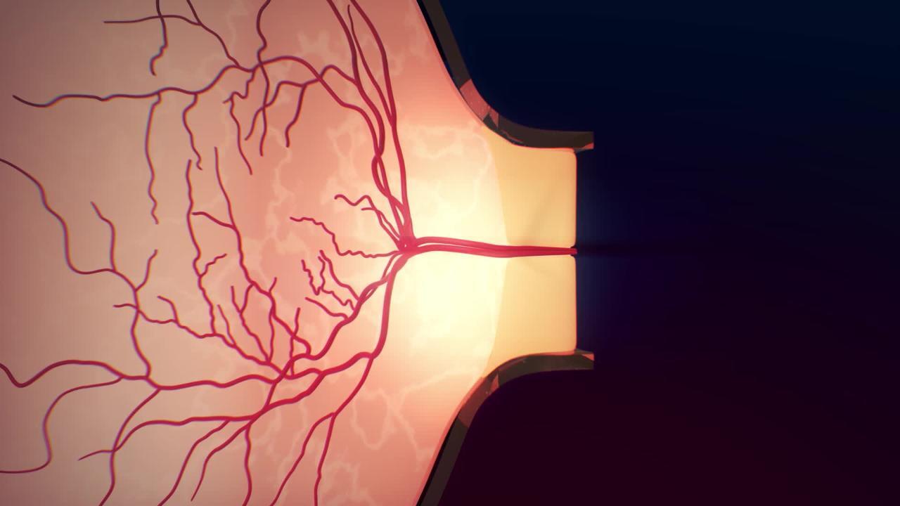
If blood pressure becomes dangerously high (called hypertensive emergency), the retinal veins can become dilated and twisted, and the optic disk (where the optic nerve meets the retina) may swell (called papilledema).
(See also Overview of Retinal Disorders.)
Diagnosis of Hypertensive Retinopathy
A doctor's examination of the eye
Using an ophthalmoscope, doctors can observe the typical appearance of the retina in people with high blood pressure. The amount of damage to the retinal blood vessels tends to correlate with the amount of damage to blood vessels in other organs affected by hypertension, such as the brain, heart, and kidneys. When blood pressure is extremely high, doctors may be able to see swelling of the optic disk (called papilledema), indicating the need for immediate treatment (see table ).
Did You Know...
|
Treatment of Hypertensive Retinopathy
Measures to treat high blood pressure
The goal of treatment for hypertensive retinopathy is to lower blood pressure long term. Rarely, when high blood pressure is severe and life threatening, treatment may be needed immediately to save vision and avoid other complications, including stroke, heart failure, kidney failure, and heart attack.
More Information
The following English-language resource may be useful. Please note that THE MANUAL is not responsible for the content of this resource.
National Eye Institute: A resource for learning about eye health (in English and Spanish) for adults and children, as well as for providing access to outreach campaigns. Simply type in the appropriate search term.


