

Rash is a common symptom, particularly during infancy. Most rashes are not serious.
Etiology of Rash in Infants and Young Children
Rashes can be caused by infection (viral, fungal, or bacterial), contact with irritants, atopy, drug hypersensitivity, other allergic reactions, inflammatory conditions, or vasculitides (see table ).

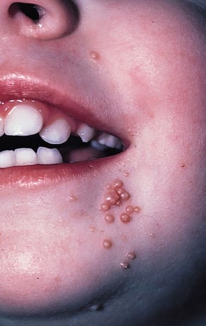
The photo shows lesions of molluscum contagiosum. Lesions are typically 1 to 5 mm, solitary or grouped, firm, painless papules. They are pearly to pink in color, dome shaped, and may be umbilicated.
The photo shows lesions of molluscum contagiosum. Lesions are typically 1 to 5 mm, solitary or grouped, firm, painless
© Springer Science+Business Media

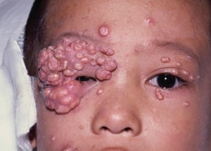
This photo shows very severe lesions on the face of a child with HIV infection. Giant molluscum indicates advanced immunodeficiency.
This photo shows very severe lesions on the face of a child with HIV infection. Giant molluscum indicates advanced immu
© Springer Science+Business Media

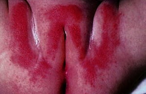
This photo shows irritant diaper dermatitis (“W-dermatitis”).
This photo shows irritant diaper dermatitis (“W-dermatitis”).
© Springer Science+Business Media

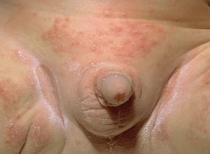
Image provided by Thomas Habif, MD.

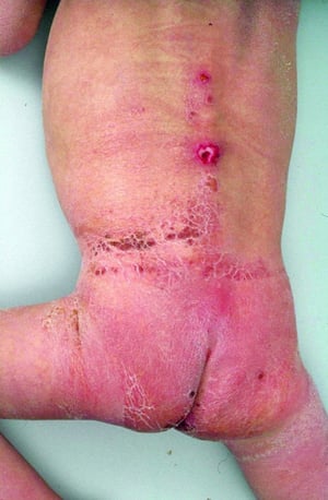
This photo shows severe diaper dermatitis due to neglect.
This photo shows severe diaper dermatitis due to neglect.
© Springer Science+Business Media

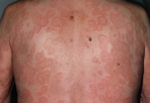
Erythema multiforme is characterized by target or iris lesions, which are annular lesions with a violaceous center and pink halo separated by a pale ring.
Erythema multiforme is characterized by target or iris lesions, which are annular lesions with a violaceous center and
DR P. MARAZZI/SCIENCE PHOTO LIBRARY

This photo shows small, pearl-colored cysts commonly seen on the face of neonates.
This photo shows small, pearl-colored cysts commonly seen on the face of neonates.
SCIENCE PHOTO LIBRARY

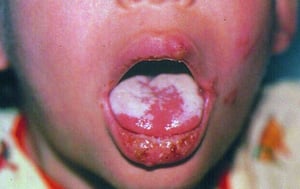
This photo shows fluffy white exudate (thrush) on the tongue of a child with HIV infection.
This photo shows fluffy white exudate (thrush) on the tongue of a child with HIV infection.
© Springer Science+Business Media

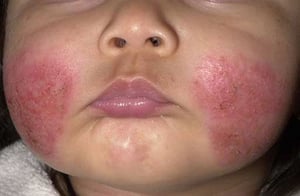
Atopic dermatitis usually develops in infancy. In the acute phase, lesions appear on the face and then spread to the neck, scalp, and extremities.
Atopic dermatitis usually develops in infancy. In the acute phase, lesions appear on the face and then spread to the ne
Image provided by Thomas Habif, MD.


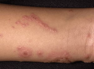
This photo shows allergic contact dermatitis on the forearm of a child after a temporary, black "henna" tattoo was applied.
This photo shows allergic contact dermatitis on the forearm of a child after a temporary, black "henna" tattoo was appl
© Springer Science+Business Media
Skin manifestations of allergic contact dermatitis range from erythema through vesiculation to edema with bullae. Changes often occur in a pattern or distribution that suggests a specific exposure. In this image, linear streaking on an extremity suggests plant contact (eg, poison ivy or poison sumac).
Skin manifestations of allergic contact dermatitis range from erythema through vesiculation to edema with bullae. Chang
Image provided by Thomas Habif, MD.


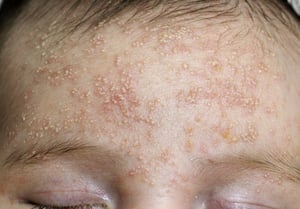
This photo shows cradle cap with thick, yellow, crusted scalp lesions and diffuse erythema.
This photo shows cradle cap with thick, yellow, crusted scalp lesions and diffuse erythema.
Biophoto Associates/SCIENCE PHOTO LIBRARY


Urticarial lesions (wheals or hives) are migratory, elevated, pruritic, reddish plaques caused by local dermal edema.
Urticarial lesions (wheals or hives) are migratory, elevated, pruritic, reddish plaques caused by local dermal edema.
Photo provided by Thomas Habif, MD.
The photo shows lesions of molluscum contagiosum. Lesions are typically 1 to 5 mm, solitary or grouped, firm, painless papules. They are pearly to pink in color, dome shaped, and may be umbilicated.
The photo shows lesions of molluscum contagiosum. Lesions are typically 1 to 5 mm, solitary or grouped, firm, painless
© Springer Science+Business Media
This photo shows very severe lesions on the face of a child with HIV infection. Giant molluscum indicates advanced immunodeficiency.
This photo shows very severe lesions on the face of a child with HIV infection. Giant molluscum indicates advanced immu
© Springer Science+Business Media
This photo shows irritant diaper dermatitis (“W-dermatitis”).
This photo shows irritant diaper dermatitis (“W-dermatitis”).
© Springer Science+Business Media
Image provided by Thomas Habif, MD.
This photo shows severe diaper dermatitis due to neglect.
This photo shows severe diaper dermatitis due to neglect.
© Springer Science+Business Media
Erythema multiforme is characterized by target or iris lesions, which are annular lesions with a violaceous center and pink halo separated by a pale ring.
Erythema multiforme is characterized by target or iris lesions, which are annular lesions with a violaceous center and
DR P. MARAZZI/SCIENCE PHOTO LIBRARY
This photo shows small, pearl-colored cysts commonly seen on the face of neonates.
This photo shows small, pearl-colored cysts commonly seen on the face of neonates.
SCIENCE PHOTO LIBRARY
This photo shows fluffy white exudate (thrush) on the tongue of a child with HIV infection.
This photo shows fluffy white exudate (thrush) on the tongue of a child with HIV infection.
© Springer Science+Business Media
Atopic dermatitis usually develops in infancy. In the acute phase, lesions appear on the face and then spread to the neck, scalp, and extremities.
Atopic dermatitis usually develops in infancy. In the acute phase, lesions appear on the face and then spread to the ne
Image provided by Thomas Habif, MD.
This photo shows allergic contact dermatitis on the forearm of a child after a temporary, black "henna" tattoo was applied.
This photo shows allergic contact dermatitis on the forearm of a child after a temporary, black "henna" tattoo was appl
© Springer Science+Business Media
Skin manifestations of allergic contact dermatitis range from erythema through vesiculation to edema with bullae. Changes often occur in a pattern or distribution that suggests a specific exposure. In this image, linear streaking on an extremity suggests plant contact (eg, poison ivy or poison sumac).
Skin manifestations of allergic contact dermatitis range from erythema through vesiculation to edema with bullae. Chang
Image provided by Thomas Habif, MD.
This photo shows cradle cap with thick, yellow, crusted scalp lesions and diffuse erythema.
This photo shows cradle cap with thick, yellow, crusted scalp lesions and diffuse erythema.
Biophoto Associates/SCIENCE PHOTO LIBRARY
Urticarial lesions (wheals or hives) are migratory, elevated, pruritic, reddish plaques caused by local dermal edema.
Urticarial lesions (wheals or hives) are migratory, elevated, pruritic, reddish plaques caused by local dermal edema.
Photo provided by Thomas Habif, MD.
Overall, the most common causes of rash in infants and young children include
Diaper rash (with or without candidal infection)
Viral exanthem
Numerous viral infections cause rash. Some (eg, chickenpox and measles, both of which are currently uncommon because of vaccination but should be considered in unvaccinated children; erythema infectiosum) have a fairly typical appearance and clinical manifestation; others are nonspecific. Cutaneous drug reactions are usually self-limited , but sometimes more serious reactions occur.
Uncommon but serious causes of rash include
Some Causes of Rash in Infants and Children
Cause | Suggestive Findings | Diagnostic Approach |
|---|---|---|
Infections | ||
Candidal infections | Beefy red rash with adjacent satellite lesions in the diaper area, including skin creases Often fluffy white plaques on the tongue or oral mucosa Sometimes history of recent antibiotic use | History and physical examination Sometimes scrapings of lesions for potassium hydroxide wet mount |
Red dots on the face, scalp, torso and proximal extremities that progress over 10–12 hours to small bumps, vesicles, and then umbilicated pustules, which form crusts Intensely itchy blisters, which may also occur on the palms, soles, scalp, and mucous membranes, as well as in the diaper area | History and physical examination | |
Confluent erythema on cheeks (slapped-cheek appearance) Sometimes fever, malaise | History and physical examination | |
Nonbullous impetigo: Painless but itchy red sore near the nose or mouth that soon leaks pus or fluid and forms a honey-colored scab Bullous impetigo: Occurs mainly in children < 2 years Painless, fluid-filled bullae—mostly on the arms, legs, and trunk, surrounded by red and itchy skin—which, after breaking, form yellow or silvery scabs | History and physical examination | |
Erythema migrans rash; an enlarging (to about 5–7 cm), erythematous lesion sometimes with central clearing or rarely purpura (2%) Often fatigue, headache, joint or body aches Usually in endemic area with risk of exposure to ticks, with or without a known tick bite | History and physical examination Sometimes serologic testing | |
Maculopapular rash beginning on the face and spreading to the trunk and extremities Often Koplik spots (white spots on buccal mucosa) Fever, cough, coryza, conjunctival injection | History and physical examination Serologic testing (for public health reasons) | |
Petechial rash, sometimes with purpura fulminans Fever, lethargy, irritability In older children, meningeal signs Tachycardia, sometimes hypotension | Gram stain and culture of blood and cerebrospinal fluid | |
Clusters of flesh-colored, umbilicated papules No itching or discomfort | History and physical examination | |
Maculopapular rash that appears suddenly after 4 or 5 days of high fever, typically as fever resolves | History and physical examination | |
Sometimes itchy rash that begins on the face and spreads downward, appears as pink or light red spots (which may merge to form evenly colored patches), and usually clears on the face as it spreads Lasts up to 3 days Often lymphadenopathy (occipital, postauricular, posterior cervical), mild fever | History and physical examination Serologic testing (for public health reasons) | |
Scarlet fever (scarlatina) | Fever, sometimes sore throat Generalized fine, red, rough-textured, blanching rash that typically appears 12–72 hours after the fever and starts on the chest, in the armpits, and on the groin Characteristic pale area around the mouth (circumoral pallor) and accentuation in the skinfolds (Pastia lines), strawberry tongue Often followed by extensive desquamation of the palms and soles, tips of fingers and toes, and groin | History and physical examination Often rapid streptococcal assay or throat culture |
Widespread areas of painful erythema that develop large, flaccid blisters, which are easily ruptured, leaving large areas of desquamation Lateral extension of blisters with gentle pressure (positive Nikolsky sign) Spares the mucous membranes Usually in children < 5 years | History and physical examination Sometimes confirmed by biopsy and/or cultures | |
Scaly, oval lesions with a slightly raised border and central clearing Mild itching | History and physical examination Sometimes scrapings of lesions for potassium hydroxide wet mount | |
Viral infection (systemic) | Maculopapular rash Often viral respiratory prodrome | History and physical examination |
Hypersensitivity reactions | ||
Atopic dermatitis (eczema) | Chronic or recurrent red, scaly patches, often in flexor creases Sometimes family history | History and physical examination |
Intensely itchy erythema, sometimes with vesicles No systemic manifestations | History and physical examination | |
Diffuse maculopapular rash History of current or recent (within 1 week) medication use | History and physical examination | |
Prodrome of fever, malaise, cough, sore throat, and conjunctivitis Painful mucosal ulcers, almost always in the mouth and lips but sometimes in the genital and anal regions Widespread areas of painful erythema that develop large, flaccid blisters, which are easily ruptured, leaving large areas of desquamation; possibly affecting the soles but usually not the scalp Lateral extension of blisters with gentle pressure (positive Nikolsky sign) Sometimes use of a causative medication (eg, sulfonamides, penicillins, antiseizure medications) | History and physical examination Sometimes biopsy | |
Well-circumscribed, pruritic, red, raised lesions With or without history of exposure to known or potential allergens | History and physical examination | |
Vasculitides | ||
Palpable purpura appearing in crops over days to weeks, typically in dependent areas (eg, legs, buttocks) Often arthritis, abdominal pain Sometimes hematuria, heme-positive stool, and/or intussusception Usually in children < 10 years | History and physical examination Sometimes skin biopsy | |
Diffuse erythematous maculopapular rash that can vary in appearance (eg, urticarial, target-like, purpuric) but never bullous or vesicular; may involve the palms and/or soles Fever (often > 39° C) for > 5 days Red, cracked lips; strawberry tongue; conjunctivitis; cervical lymphadenopathy Edema of hands and feet Later desquamation of fingers and toes extending to palms and soles | Clinical criteria Testing to exclude other disorders | |
Other | ||
Red and yellow scaling on the scalp (cradle cap) and sometimes in skinfolds | History and physical examination | |
Diaper rash (noncandidal) | Bright red rash in the diaper area, sparing creases | History and physical examination |
Petechial rash, pallor Usually during or after infectious colitis manifesting with abdominal pain, vomiting, and bloody diarrhea Oliguria or anuria Hypertension | Complete blood count with platelets and peripheral smear to check for evidence of microangiopathic anemia and thrombocytopenia Renal function tests Stool testing (Shiga toxin assay or specific culture for E. coli O157:H7) | |
Small pearly cysts on a neonate's face | History and physical examination | |
Pink-red blotches, symmetrically arranged and starting on the extremities, then evolving into the classic target-like lesion with a pink-red ring around a pale center Sometimes oral mucosal lesions, pruritis | History and physical examination | |
Miliaria (heat rash) | Small red bumps or occasionally small blisters Most common in very young children but can occur at any age, particularly during hot and humid weather | History and physical examination |
Erythema toxicum | Flat red splotches (usually with a white, pimple-like bump in the middle), which appear in up to half of all babies Rarely appears after 5 days of age and is usually gone in 7–14 days | History and physical examination |
Neonatal acne | Red bumps, sometimes with white dots in the center on a neonate's face Usually occurs between 2 and 4 weeks after birth but may appear up to 4 months after birth and can last for 12–18 months | History and physical examination |
Sometimes upper respiratory infection prodrome Typically begins as a single, pruritic 2- to 10-cm oval red herald patch on the trunk or proximal limbs 7–14 days after the herald patch, appearance of large patches of pink or red, flaky, oval-shaped rash on the torso, sometimes in a characteristic Christmas tree–like distribution | History and physical examination | |
* This cause is currently uncommon because of vaccination but should be considered in unvaccinated children. | ||
Evaluation of Rash in Infants and Young Children
History
History of present illness is focused on the time course of illness, particularly the relationship between the rash and other symptoms.
Review of systems is focused on symptoms of causative disorders, including gastrointestinal symptoms (suggesting immunoglobulin A–associated vasculitis [formerly called Henoch-Schönlein purpura] or hemolytic-uremic syndrome), joint symptoms (suggesting immunoglobulin A–associated vasculitis or Lyme disease), headache or neurologic symptoms (suggesting meningitis or Lyme disease).
Past medical history should note any medications recently used, particularly antibiotics and antiseizure medications. Family history of atopy is noted.
Physical examination
Examination begins with a review of vital signs, particularly to check for fever. At initial observation, the child is assessed for signs of lethargy, irritability, or distress. A full physical examination is done, with particular attention to the characteristics of the skin lesions, including the presence of vesicles, bullae, petechiae, purpura, or urticaria and mucosal involvement. Children are evaluated for meningeal signs (neck stiffness, Kernig and Brudzinski signs), although these signs are often absent in children < 2 years.
Red flags
The following findings are of particular concern:
Bullae or skin sloughing
Diarrhea and/or abdominal pain
Fever and inconsolability or extreme irritability
Mucosal inflammation
Petechiae and/or purpura
Urticaria with respiratory distress
Interpretation of findings
Well-appearing children without systemic symptoms or signs are unlikely to have a dangerous disorder. The appearance of the rash typically narrows the differential diagnosis. The associated symptoms and signs help identify patients with a serious disorder and often suggest the diagnosis (see table ).
Bullae and/or sloughing suggest staphylococcal scalded skin syndrome or Stevens-Johnson syndrome and are considered dermatologic emergencies. Conjunctival inflammation may occur in Kawasaki disease, measles, staphylococcal scalded skin syndrome, and Stevens-Johnson syndrome. Any child presenting with fever and petechiae or purpura must be evaluated carefully for the possibility of meningococcemia. Bloody diarrhea with pallor and petechiae should raise concern about the possibility of hemolytic uremic syndrome. Fever for > 5 days with evidence of mucosal inflammation and rash should prompt consideration of and further evaluation for Kawasaki disease.
Testing
For most children, the history and physical examination are sufficient for diagnosis. Testing is targeted at potential life threats; it includes Gram stain and cultures of blood and cerebrospinal fluid for meningococcemia; complete blood count, renal function tests, and stool tests for hemolytic uremic syndrome.
Treatment of Rash in Infants and Young Children
Treatment of rash is directed at the cause (eg, antifungal cream for candidal infection).
For diaper rash, the goal is to keep the diaper area clean and dry, primarily by changing diapers more frequently and gently washing the area with mild soap and water. Sometimes a barrier ointment containing zinc oxide or vitamins A and D may help.For diaper rash, the goal is to keep the diaper area clean and dry, primarily by changing diapers more frequently and gently washing the area with mild soap and water. Sometimes a barrier ointment containing zinc oxide or vitamins A and D may help.
Pruritus in infants and children can be lessened by oral antihistamines (eg, diphenhydramine, hydroxyzine, cetirizine, loratadine).Pruritus in infants and children can be lessened by oral antihistamines (eg, diphenhydramine, hydroxyzine, cetirizine, loratadine).
Some common adverse effects of antihistamines include dry mouth, drowsiness, dizziness, nausea and vomiting, restlessness or moodiness (in some children), urinary hesitancy, blurred vision, and confusion.
Key Points
Most rashes in children are benign.
For most rashes in infants and children, the history and physical examination are sufficient for diagnosis.
Children with rash due to serious illness typically have systemic manifestations of disease.
Drug Information for the Topic


