

Patellar dislocations are common and almost always lateral. Diagnosis is clinical; radiographs are performed to exclude fracture. Treatment is reduction and immobilization.
Patellar dislocation is distinct from knee dislocation, which is a much more serious injury.
Most patients are adolescent females and have an underlying chronic patellofemoral abnormality. Many dislocations spontaneously reduce before medical evaluation.
Usually, patellar dislocation occurs when people suddenly change direction or twist the knee or when force is applied to the knee (as may occur in soccer, gymnastics, or baseball when swinging a bat).
Associated injuries include:
Osteochondral fracture of the patella or lateral femoral condyle
Complications can include:
In patients with patellofemoral abnormalities, recurrent dislocation or subluxation
(See Overview of Dislocations.)
Diagnosis of Patellar Dislocations
Radiographs to exclude fracture
A patellar dislocation, unless spontaneously reduced, is clinically obvious; ie, the patella is visibly and palpably displaced laterally, and the patient holds the knee in a slightly flexed position and is unwilling to straighten it. If the dislocation has spontaneously reduced, hemarthrosis is often present, and the peripatellar area is usually tender.

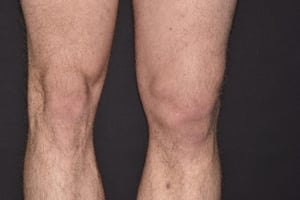
Close-up comparing the normal right knee and swollen left knee of a male patient following dislocation of the kneecap (patella) as a result of an injury.
Close-up comparing the normal right knee and swollen left knee of a male patient following dislocation of the kneecap (
DR P. MARAZZI/SCIENCE PHOTO LIBRARY

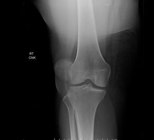
This anteroposterior view of the knee shows a patellar dislocation, characterized by extreme lateral displacement of the patella, outside its normal location between the femoral condyles.
This anteroposterior view of the knee shows a patellar dislocation, characterized by extreme lateral displacement of th
Image courtesy of Danielle Campagne, MD.
Close-up comparing the normal right knee and swollen left knee of a male patient following dislocation of the kneecap (patella) as a result of an injury.
Close-up comparing the normal right knee and swollen left knee of a male patient following dislocation of the kneecap (
DR P. MARAZZI/SCIENCE PHOTO LIBRARY
This anteroposterior view of the knee shows a patellar dislocation, characterized by extreme lateral displacement of the patella, outside its normal location between the femoral condyles.
This anteroposterior view of the knee shows a patellar dislocation, characterized by extreme lateral displacement of th
Image courtesy of Danielle Campagne, MD.
Anteroposterior and lateral knee radiographs and patellar views are performed to exclude fracture, even if the dislocation has obviously reduced.
Treatment of Patellar Dislocations
Reduction
Immobilization
Immediate treatment of patellar dislocations is reduction; most patients do not require sedation or analgesia. Reduction is performed with the patient's hip flexed. Then clinicians gently move the patella medially while simultaneously extending the knee. When the patella is reduced, a palpable clunk is usually evident and the deformity resolves. (See also How To Reduce a Lateral Patellar Dislocation.)
Immediately after reduction, the knee can be checked for stability by moving it through its range of motion (flexion and extension). If the knee is stable, treatment consists of crutches and an elastic wrap. If the knee is unstable, a knee immobilizer is needed.


Patients with osteochondral injury or recurrent instability may require a referral to an orthopedic surgeon for outpatient surgery.
Key Points
Patellar dislocations, which are common, are distinct from knee dislocations, which are much more serious.
Patellar dislocations occur most often in adolescent females who have an underlying chronic patellofemoral abnormality.
A patellar dislocation, unless spontaneously reduced, is usually clinically obvious; perform radiographs to exclude fracture.
Reduce the dislocation and immobilize the knee.
If patients have an osteochondral injury or recurrent instability, refer them to an orthopedic surgeon.


