

Most elbow dislocations are posterior and usually result from a fall on an extended arm.
Posterior elbow dislocations are common; it is the second most common joint dislocation after shoulder dislocations. Associated injuries may include:
Fractures
Injuries to the ulnar or median nerve
Possibly injury to the brachial artery

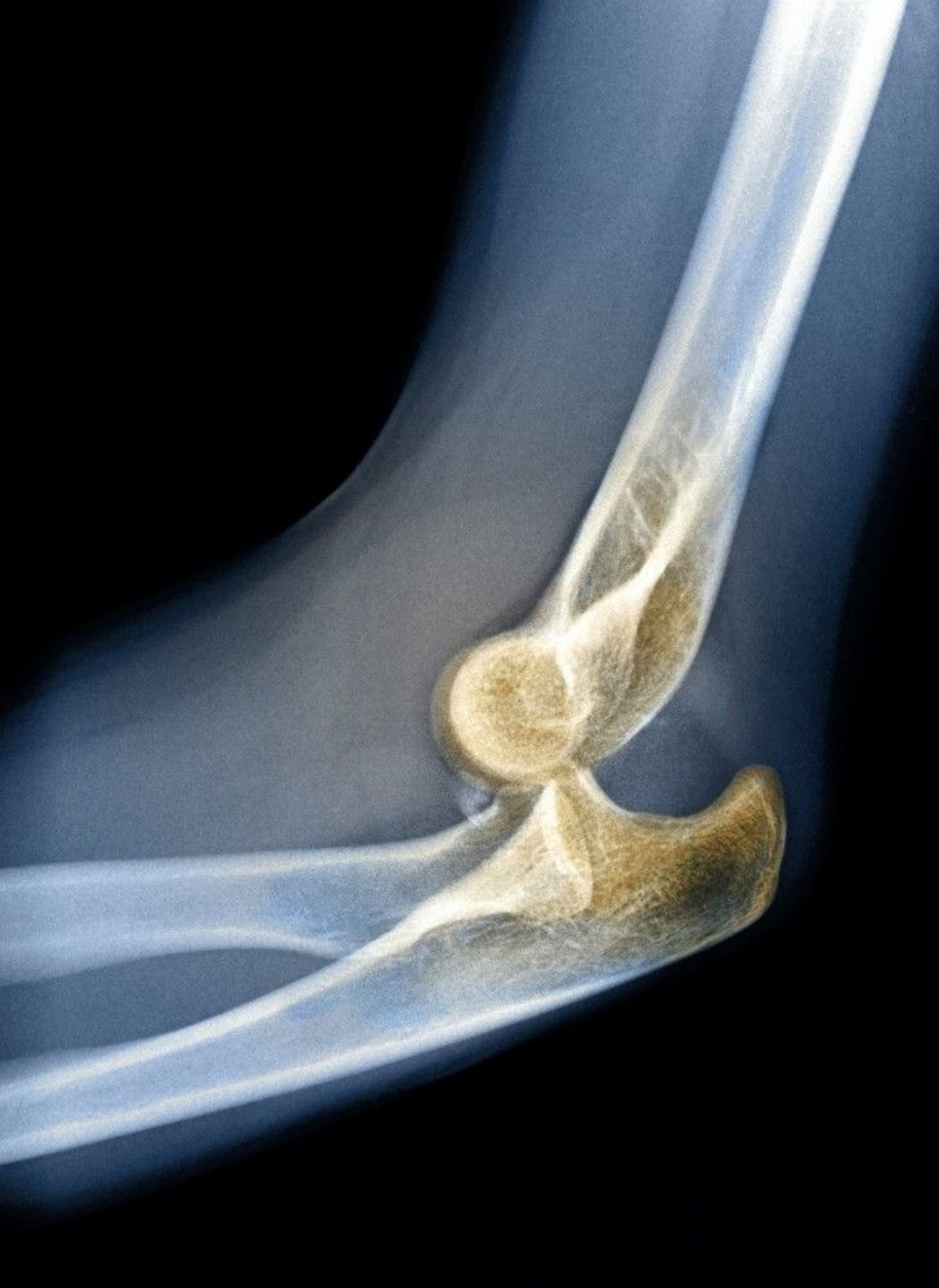
DU CANE MEDICAL IMAGING LTD/SCIENCE PHOTO LIBRARY
The joint is usually flexed approximately 45°, and the olecranon is prominent and posterior to the humeral epicondyles; however, these anatomic relationships may be difficult to determine because of swelling. Classically, patients with an elbow dislocation present with a shortened forearm and a very prominent olecranon.
Radiographs are diagnostic.
(See Overview of Dislocations.)
Treatment of Elbow Dislocations
Traction to reduce the elbow joint, usually with procedural sedation
For elbow dislocations, reduction is usually with sustained, gentle traction and correction of deformity after patients are sedated and given analgesics (For detailed instructions, see How To Reduce a Posterior Elbow Dislocation). The following technique is commonly used:
With the patient supine, the clinician flexes the elbow to approximately 90° and supinates the forearm.
An assistant stabilizes the upper arm against the stretcher.
The clinician grasps the wrist and applies slow, steady axial traction to the forearm while keeping the elbow flexed and the forearm supinated.
Traction is maintained until the dislocation is reduced.
After reduction, the clinician checks the elbow for stability by fully flexing and extending the elbow while pronating and supinating the forearm. These movements should be easy after reduction. After reduction, radiographs should be performed to make sure no fractures were missed.
The joint is immobilized. If the joint is stable, immobilization is with a sling, and range-of-motion exercises begin 1 week later. If the joint has laxity, immobilization is with a splint; the patient follows up with an orthopedic surgeon, who determines further management, including when to begin range-of-motion exercises.
Key Points
Many patients with an elbow dislocation present with a shortened forearm and a very prominent olecranon; the position of the bones may be difficult to determine because of swelling.
Perform radiographs to diagnose a dislocated elbow.
Apply gentle, sustained traction to reduce the joint after patients are sedated and given analgesics.
After reduction, check the joint for stability, perform radiographs to check for fractures, and immobilize the joint, especially if laxity is present.


