

Conventional radiography involves the use of x-rays; the term “plain x-rays” is sometimes used to distinguish x-rays used alone from x-rays combined with other techniques (eg, CT).
For conventional radiography, an x-ray beam is generated and passed through a patient to a piece of film or a radiation detector, producing an image. Different soft tissues attenuate x-ray photons differently, depending on tissue density; the denser the tissue, the whiter (more radiopaque) the image. The range of densities, from most to least dense, is represented by metal (white, or radiopaque), bone cortex (less white), muscle and fluid (gray), fat (darker gray), and air or gas (black, or radiolucent).
Uses of Conventional Radiography
Radiography is the most readily available imaging method. Typically, it is the first imaging method indicated to evaluate the extremities, chest, and sometimes the spine and abdomen. These areas contain important structures with densities that differ from those of adjacent tissues. For example, radiography is a first-line test for detecting the following:
Fractures: White bone is well seen because it is adjacent to gray soft tissues.
Pneumonia: Inflammatory exudate that fills the lungs is well seen because it contrasts with adjacent, more radiolucent air spaces.
Intestinal obstruction: Dilated, air-filled loops of intestine are well seen amidst the surrounding soft tissue.

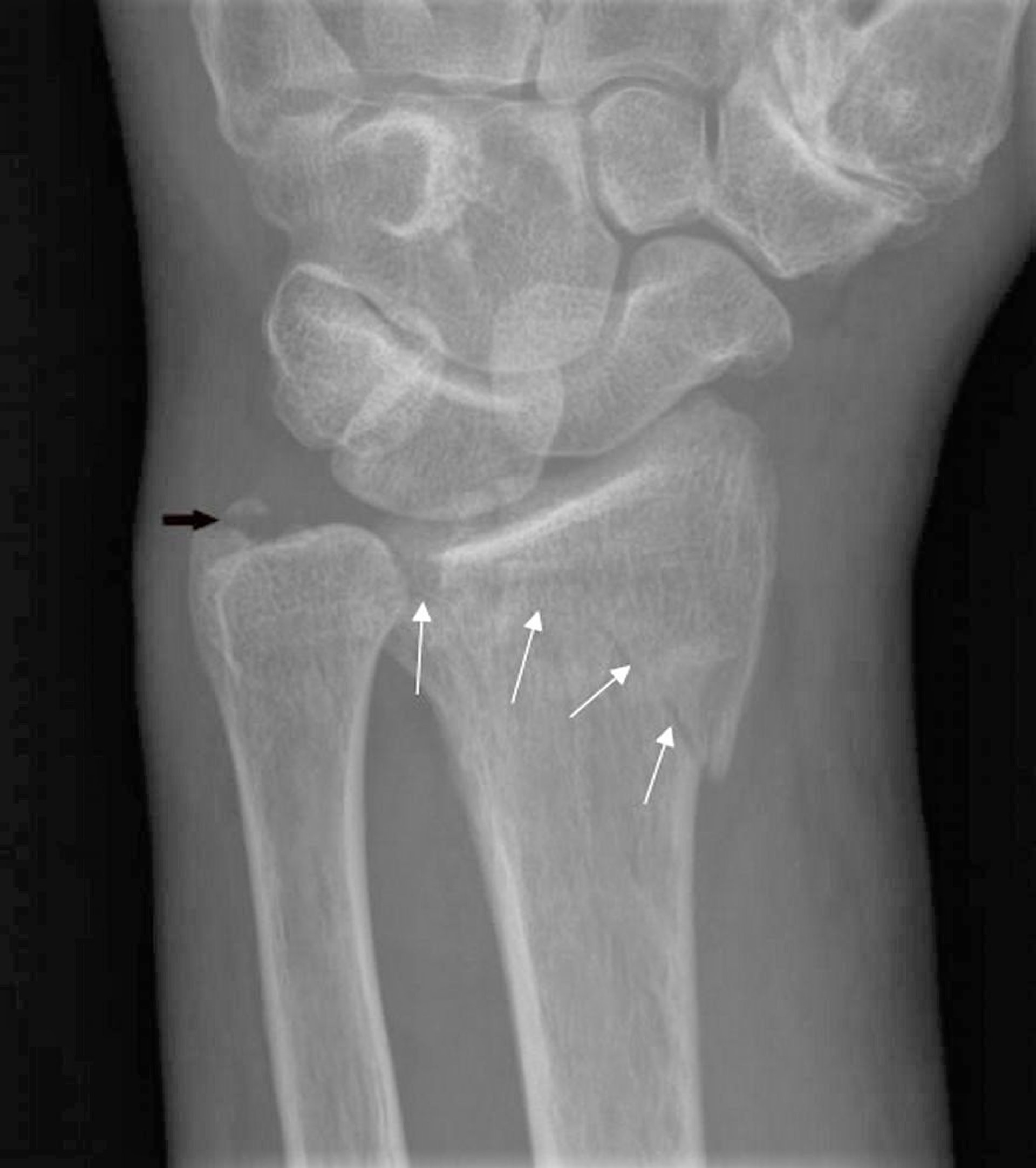
Anteroposterior radiograph of the left wrist shows a minimally displaced intra-articular fracture of the distal radius (white arrows) and ulnar styloid avulsion (black arrow).
Image courtesy of Hakan Ilaslan, MD.

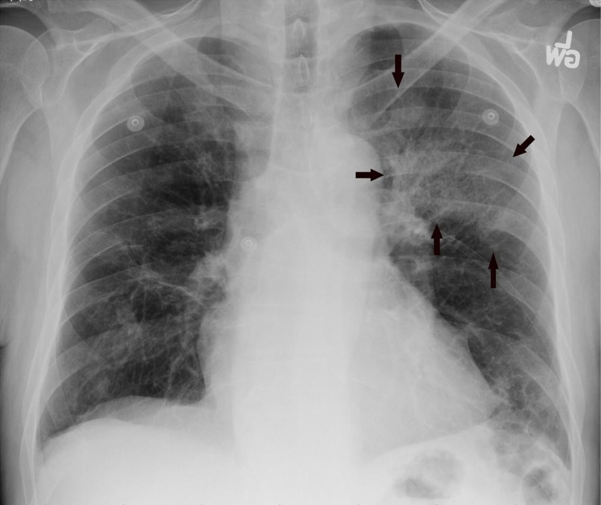
Posteroanterior radiograph of the chest shows left upper lobe infiltrate consistent with pneumonia (arrows).
Image courtesy of Hakan Ilaslan, MD.
Variations of Conventional Radiography
Contrast studies
When the density of adjacent tissues is similar, a radiopaque contrast agent is often added to one tissue or structure to differentiate it from its surroundings. Structures typically requiring a contrast agent include blood vessels (for angiography) and the lumina of the gastrointestinal, biliary, and genitourinary tracts. Gas may be used to distend the lower gastrointestinal tract and make it visible.
Other imaging tests (eg, CT, MRI) have largely replaced contrast studies because their tomographic images provide better anatomic localization of an abnormality. Endoscopic procedures have largely replaced barium contrast studies of the esophagus, stomach, and upper intestinal tract.
Fluoroscopy
A continuous x-ray beam is used to produce real-time images of moving structures or objects. Fluoroscopy is most often used:
With contrast agents (eg, in swallowing studies or coronary artery catheterization)
During medical procedures to guide placement of a cardiac lead, catheter, or needle (eg, in electrophysiologic testing or percutaneous coronary interventions)
Fluoroscopy can also be used in real time to detect motion of the diaphragm and of bones and joints (eg, to assess the stability of musculoskeletal injuries).
Disadvantages of Conventional Radiography
Diagnostic accuracy is limited in many situations. Other imaging tests may have advantages, such as providing better detail or being safer or faster.
Intestinal contrast agents such as barium and gastrografin (an iodine-based oral contrast agent), if used, have disadvantages (see Disadvantages of CT), and IV contrast agents have risks.
Fluoroscopy may involve high doses of radiation (see Risks of Medical Radiation).


