

Diaphragmatic hernia is protrusion of abdominal contents into the thorax through a defect in the diaphragm. Pulmonary hypoplasia may cause pulmonary hypertension. Diagnosis is by fetal ultrasound, chest radiograph, or other advanced imaging. Treatment is surgical repair and long-term management.
(See also Overview of Congenital Gastrointestinal Anomalies.)
Diaphragmatic hernia, also called congenital diaphragmatic hernia, occurs in the posterolateral portion of the diaphragm (Bochdalek hernia) in 95% of cases and is on the left side in 85% of cases; in 2% of cases it is bilateral (1). The estimated birth prevalence is approximately 1 in 4000 live births (2). Anterior hernias (Morgagni hernia) are far less common (5% of cases). Other congenital anomalies, particularly congenital heart disease, are present in approximately 50% of cases (3).
Loops of small and large bowel, stomach, liver, and spleen may protrude into the hemithorax on the involved side. If the hernia is large and the amount of herniated abdominal contents is substantial, the lung on the affected side is hypoplastic; however, the lung on the opposite side can also be adversely affected. Other pulmonary consequences include underdevelopment of the pulmonary vasculature, resulting in an elevation of pulmonary vascular resistance and hence pulmonary hypertension. Pulmonary hypertension may lead to right-to-left shunting at the level of the foramen ovale or through a patent ductus arteriosus; both pulmonary hypoplasia and pulmonary hypertension prevent adequate oxygenation, even with oxygen supplementation or mechanical ventilation. Additionally, alterations in myocardial contractility add to the complexity of management and contribute to mortality and morbidity (4).
General references
1. Deprest JA, Nicolaides KH, Benachi A, et al. Randomized Trial of Fetal Surgery for Severe Left Diaphragmatic Hernia. N Engl J Med. 2021;385(2):107-118. doi:10.1056/NEJMoa2027030
2. Paoletti M, Raffler G, Gaffi MS, Antounians L, Lauriti G, Zani A. Prevalence and risk factors for congenital diaphragmatic hernia: A global view. J Pediatr Surg. 2020;55(11):2297-2307. doi:10.1016/j.jpedsurg.2020.06.022
3. Longoni M, Pober BR, High FA. Congenital Diaphragmatic Hernia Overview. 2006 Feb 1 [Updated 2020 Nov 5]. In: Adam MP, Feldman J, Mirzaa GM, et al., editors. GeneReviews® [Internet]. Seattle (WA): University of Washington, Seattle; 1993-2025.
4. Zani A, Chung WK, Deprest J, et al. Congenital diaphragmatic hernia. Nat Rev Dis Primers. 2022;8(1):37. Published 2022 Jun 1. doi:10.1038/s41572-022-00362-w
Symptoms and Signs of Diaphragmatic Hernia
Respiratory distress typically occurs in the first several hours after birth and occurs immediately after delivery in severe cases. After delivery, as the neonate cries and swallows air, the stomach and the loops of intestine quickly fill with air and rapidly enlarge, causing acute respiratory compromise as the heart and mediastinal structures are pushed to the right (with the most frequent left-sided hernia), compressing the more normal right lung. A scaphoid abdomen (due to displacement of abdominal viscera into the chest) is likely. Bowel sounds (and an absence of breath sounds) may be heard over the involved hemithorax.
In less severe cases, mild respiratory difficulty develops a few hours or days later as abdominal contents progressively herniate through a smaller diaphragmatic defect.
Rarely, presentation is delayed until later in childhood, sometimes after a bout of infectious enteritis, which causes sudden herniation of bowel into the chest.
Diagnosis of Diaphragmatic Hernia
Sometimes, by prenatal ultrasound or fetal MRI
Chest radiograph, chest CT and sometimes MRI
Echocardiogram
In some cases, diaphragmatic hernia can be diagnosed by prenatal ultrasound (1), with additional risk stratification available based on fetal MRI (2).
In the delivery room, diaphragmatic hernia may be suspected when neonates with respiratory distress have a scaphoid abdomen.
After delivery, diagnosis is by chest radiograph showing the stomach and intestine protruding into the chest. In a large defect, there are numerous air-filled loops of intestine filling the hemithorax and contralateral displacement of the heart and mediastinal structures. If the radiograph is taken immediately after delivery before the neonate has swallowed air, the abdominal contents appear as an opaque airless mass in the hemithorax.
Cross-sectional imaging including CT and MRI is used to evaluate the location of the defect, identify the specific abdominal contents in the thorax, approximate lung volumes, plan for surgery, and in some neonates evaluate for other congenital defects (3). Echocardiogram is performed to screen for associated congenital heart disease, particularly if extracorporeal membrane oxygenation is being considered.

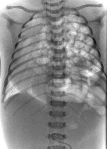
This radiograph shows congenital diaphragmatic hernia in a neonate. Loops of bowel are protruding into the left chest (right side of radiograph).


In this contrast study, stomach and bowel are seen in the left chest.
ZEPHYR/SCIENCE PHOTO LIBRARY
Diagnosis references
1. Cordier AG, Russo FM, Deprest J, et al. Prenatal diagnosis, imaging, and prognosis in congenital diaphragmatic hernia. Semin Perinatol. 2020;44(1):51163. doi:10.1053/j.semperi.2019.07.002
2. Zani A, Chung WK, Deprest J, et al. Congenital diaphragmatic hernia. Nat Rev Dis Primers. 2022;8(1):37. Published 2022 Jun 1. doi:10.1038/s41572-022-00362-w
3. Expert Panel on Gastrointestinal Imaging, Garcia EM, Pietryga JA, et al. ACR Appropriateness Criteria® Hernia. J Am Coll Radiol. 2022;19(11S):S329-S340. doi:10.1016/j.jacr.2022.09.016
Treatment of Diaphragmatic Hernia
Sometimes fetal surgery
Multidisciplinary delivery planning and critical care management
Sometimes extracorporeal membrane oxygenation (ECMO)
Surgical repair
If a diaphragmatic hernia is diagnosed in a fetus, delivery at a pediatric center with ECMO capability is recommended because of the inherent transport challenges. Fetoscopic endoluminal tracheal occlusion (FETO) may be performed in select fetuses before delivery (between approximately 27 to 34 weeks gestational age) to induce lung growth; this procedure is associated with a survival benefit in severe cases but has significant risks (1).
If diaphragmatic hernia is suspected in a neonate with respiratory distress, the neonate should be immediately endotracheally intubated and ventilated in the delivery room. Bag-and-mask ventilation should be avoided because it may fill the displaced intrathoracic viscera with air and worsen respiratory compromise. Continuous nasogastric suction with a double-lumen nasogastric tube prevents swallowed air from progressing through the gastrointestinal tract and causing further lung compression.
Preoperative critical care involves early evaluation for and management of pulmonary hypertension with inhaled nitric oxide and other agents (Preoperative critical care involves early evaluation for and management of pulmonary hypertension with inhaled nitric oxide and other agents (2, 3). The infant's acid-base status, hemodynamics, oxygenation, and ventilation are optimized. Adrenal insufficiency resulting in catecholamine-resistant hypotension is common (4). Conventional or alternative ventilatory strategies, such as high-frequency oscillating ventilation, may be used. ECMO is a commonly used as a planned approach for higher risk infants with more severe pulmonary hypoplasia or as a rescue approach for infants with refractory hypoxemia or combined cardiorespiratory failure.
Surgery is required to replace the abdominal contents into the abdomen and close the diaphragmatic defect.
Long-term management involves ongoing and multidisciplinary care of patients who with pulmonary hypertension, sequelae of pulmonary hypoplasia, gastroesophageal reflux or other gastrointestinal dysfunction, heart failure, or neurodevelopmental abnormalities.
Pearls & Pitfalls
|
Treatment references
1. Zani A, Chung WK, Deprest J, et al. Congenital diaphragmatic hernia. Nat Rev Dis Primers. 2022;8(1):37. Published 2022 Jun 1. doi:10.1038/s41572-022-00362-w
2. Puligandla P, Skarsgard E, Baird R, et al. Diagnosis and management of congenital diaphragmatic hernia: a 2023 update from the Canadian Congenital Diaphragmatic Hernia Collaborative. Arch Dis Child Fetal Neonatal Ed. 2024;109(3):239-252. Published 2024 Apr 18. doi:10.1136/archdischild-2023-325865
3. Snoek KG, Reiss IK, Greenough A, et al. Standardized Postnatal Management of Infants with Congenital Diaphragmatic Hernia in Europe: The CDH EURO Consortium Consensus - 2015 Update. Neonatology. 2016;110(1):66-74. doi:10.1159/000444210
4. Kamath BD, Fashaw L, Kinsella JP. Adrenal insufficiency in newborns with congenital diaphragmatic hernia. J Pediatr. 2010;156(3):495-497.e1. doi:10.1016/j.jpeds.2009.10.044
Key Points
A congenital diaphragmatic hernia can allow abdominal contents to enter the chest cavity, compressing the lung and causing neonatal respiratory distress.
Pulmonary hypoplasia and pulmonary hypertension are common.
Diagnosis is by prenatal imaging and by postnatal chest radiograph and cross-sectional imaging.
Treat with endotracheal intubation followed by surgical repair; often, extracorporeal membrane oxygenation is necessary.


