

Most ankle dislocations are fracture-dislocations. Reduction uses traction-countertraction to disengage the talus from the distal tibia, followed by repositioning of the talar dome into the joint mortice and splinting to stabilize the reduction until definitive orthopedic treatment. Procedural sedation and analgesia (PSA) is usually required.
(See also Overview of Dislocations and Ankle Fractures.)
Indications for Ankle Dislocation Reduction
Dislocation or fracture-dislocation of the ankle
Most ankle dislocations are posterior or posteromedial and are fracture-dislocations associated with malleolar, distal fibular, and posterior marginal tibial fractures. In a posterior dislocation, the talus is displaced posteriorly from the tibia and fibula.
Reduction of a closed ankle dislocation or fracture-dislocation should be attempted soon after the diagnosis is made. An associated neurovascular deficit or a fracture-dislocation with skin tenting that threatens skin penetration warrants immediate reduction.
Open dislocations require surgery, but closed reduction techniques and splinting should be performed as interim treatment if the orthopedic surgeon is unavailable and a neurovascular deficit is present.
Contraindications to Ankle Dislocation Reduction
There are no contraindications to attempting closed reduction of ankle dislocations, even those awaiting orthopedic evaluation and treatment. However, open dislocations without vascular compromise—if surgery is impending—may be better managed with thorough irrigation in the operating room (rather than irrigation in the emergency department) before the reduction.
Complications of Ankle Dislocation Reduction
Compartment syndrome (due to soft tissue injury and swelling from the initial trauma) and articular damage, which increase with increasing time to reduction. This complication is from the injury itself, but timely reduction may help prevent it.
Neurovascular injury (uncommon) due to either the injury or the reduction procedure
Most complications are the result of the fracture-dislocation itself.
Equipment for Ankle Dislocation Reduction
Materials and personnel required for procedural sedation and analgesia (PSA)
For intra-articular analgesia: anesthetic (eg, 5 to 10 mL of 1% lidocaine, 10-mL syringe, 5 cm (2-inch) 20-gauge needle), antiseptic solution (eg, chlorhexidine, povidone iodine), gauze padsFor intra-articular analgesia: anesthetic (eg, 5 to 10 mL of 1% lidocaine, 10-mL syringe, 5 cm (2-inch) 20-gauge needle), antiseptic solution (eg, chlorhexidine, povidone iodine), gauze pads
Pillow
Short leg splint (stockinette, cotton padding wrap, splint material [posterior, 3-sided splint], elastic bandage)
Additional Considerations for Ankle Dislocation Reduction
Radiographs should be performed before reduction of ankle dislocations unless neurovascular deficits are present, but typically radiographs can be performed in the time that it takes to get the supplies together for PSA and reduction.
Intravenous analgesia is preferably given prior to radiographs.
Intra-articular or regional anesthesia may be sufficient in some cases.
Lateral dislocations should not be reduced without orthopedic involvement unless vascularity is compromised or the patient must be transported to the orthopedic surgeon.
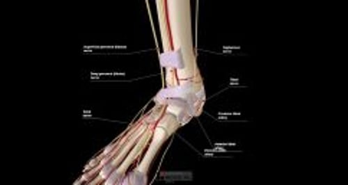
Relevant Anatomy for Ankle Dislocation Reduction
Ankle ligaments are strong and ankle dislocations are high-energy injuries that usually involve fractures and ligament ruptures. Associated fractures include those of the malleoli, fibula, or tibial margins.
Anterior dislocations may disrupt the dorsalis pedis artery.
Malleolar or distal fibular fractures generally accompany lateral dislocations.
Positioning for Ankle Dislocation Reduction
Place the patient supine, with the affected foot at the end of the stretcher and the knee in slight flexion.
Step-by-Step Description of Ankle Dislocation Reduction
Neurovascular examination
Perform a pre-procedure neurovascular examination of the foot and ankle, including posterior tibial and dorsalis pedis pulses, capillary refill time (normally < 2 seconds), and sensation of the foot's plantar surface (tibial nerve), dorsal surface (peroneal nerves), lateral surface (sural nerve), and medial surface (saphenous nerve).
Analgesia
Administer procedural sedation and analgesia (PSA).
If using intra-articular anesthesia, first clean the anteromedial ankle area with antiseptic solution. Then, insert the needle as when performing arthrocentesis, perpendicularly to the skin just distal to the tibia, if possible anterior to the medial malleolus and lateral to the tibialis anterior tendon. Apply back pressure on the syringe plunger, and advance the needle posteriorly until synovial fluid is aspirated. If any blood is aspirated from the joint, hold the needle hub motionless, switch to an empty syringe, aspirate all of the blood, and re-attach the anesthetic syringe. Then inject 5 to 10 mL of anesthetic solution. Wait for analgesia to occur (up to 15 to 20 minutes) before proceeding.
Reduce the ankle dislocation
Place a pillow behind the knee of the affected leg, to flex the hip and the knee.
Have one assistant grasp the calf with both hands, ready to pull cephalad (countertraction).
Have a second assistant grasp the ankle with one hand (to stabilize lower leg).
Grasp the foot, with one hand at the heel and the other hand at the forefoot.
For a posterior dislocation:
First free the talus from the distal tibia: Slightly plantarflex the foot and distract the heel axially (ie, pull it away) from the tibia, with the first assistant providing axial countertraction to the calf.
Next, while maintaining axial distraction of the heel, and with the second assistant applying a counterforce to the anterior ankle, dorsiflex the foot, to reposition the talar dome anteriorly into the joint mortice.
For an anterior dislocation:
First dorsiflex the foot to distract the talus from the tibia.
Apply axial traction and then push the foot directly backward while an assistant applies countertraction to the posterior part of the leg.
For a lateral dislocation:
Distract the heel axially from the tibia, then move the foot medially and dorsiflex it.
For all dislocations:
Successful reduction may be accompanied by a perceptible "clunk."
Aftercare for Ankle Dislocation Reduction
Successful reduction is preliminarily confirmed by visible restoration of a normal calcaneal contour and by decreased pain.
Perform a post-procedure neurovascular examination. A post-procedure neurovascular deficit warrants emergency orthopedic evaluation.
Apply a long leg posterior splint with 90° of foot dorsiflexion along with a stirrup splint to provide additional stability.
Perform post-procedure radiographs to confirm proper reduction and identify any previously unidentified fractures.
Keep the limb elevated and check for neurovascular deficits and development of compartment syndrome in consultation with an orthopedic surgeon.
Arrange orthopedic follow-up.
Warnings and Common Errors for Ankle Dislocation Reduction
Ankle joint fracture-dislocations with 2 or more areas of fracture are inherently unstable; exercise caution when transporting a patient with an ankle that is still dislocated because neurovascular compromise may develop en route.


