

An ophthalmic nerve block anesthetizes the ipsilateral forehead, frontal scalp, and sometimes the upper eyelid.
(See also Local anesthesia for laceration treatment.)
Indications for Ophthalmic Nerve Block
Laceration or other surgically treated lesion of the frontal scalp, forehead, eyebrow, or upper eyelid
A nerve block has advantages over local anesthetic infiltration when accurate approximation of wound edges is important (eg, in facial skin repair), because a nerve block does not distort the tissue as does local infiltration.
Contraindications to Ophthalmic Nerve Block
Absolute contraindications
History of allergy to the anesthetic agent
Absence of anatomic landmarks needed to guide needle insertion (eg, due to trauma)
Relative contraindications
Infection in the path of needle insertion: Use procedural sedation or a different means of anesthesia.
Coagulopathy*: When feasible, correct prior to procedure, or use a different means of analgesia.
* Anticoagulant medications (eg, for atrial fibrillation) increase the risk of bleeding with nerve blocks, but this must be balanced against the increased risk of thrombosis (eg, stroke) if anticoagulation is reversed. Discuss any contemplated reversal with the clinician managing the patient's anticoagulation and then with the patient.
Complications of Ophthalmic Nerve Block
Adverse reaction to the anesthetic (see Local anesthesia for laceration treatment)
Toxicity due to anesthetic overdose (eg, seizure, cardiac arrhythmias) or sympathomimetic effects due to epinephrine (if using an anesthetic-epinephrine mixture)Toxicity due to anesthetic overdose (eg, seizure, cardiac arrhythmias) or sympathomimetic effects due to epinephrine (if using an anesthetic-epinephrine mixture)
Intravascular injection of anesthetic or epinephrine Intravascular injection of anesthetic or epinephrine
Hematoma
Neuritis
Spread of infection, by passing the needle through an infected area
Most complications result from inaccurate needle placement.
Equipment for Ophthalmic Nerve Block
Gloves (sterile gloves are not required)
Personal protective equipment as indicated (eg, face mask, safety glasses or face shield, cap and gown)
Antiseptic solution (eg, chlorhexidine, povidone-iodine, alcohol)Antiseptic solution (eg, chlorhexidine, povidone-iodine, alcohol)
Injectable local anesthetic* such as lidocaine 2% with epinephrine† 1:100,000 or, for longer-duration anesthesia, bupivacaine 0.5% with epinephrine† 1:200,000 Injectable local anesthetic* such as lidocaine 2% with epinephrine† 1:100,000 or, for longer-duration anesthesia, bupivacaine 0.5% with epinephrine† 1:200,000
Syringe (eg, 3 mL) and needle (eg, 25 or 27 gauge) for anesthetic injection
* Local anesthetics are discussed in Local anesthesia for laceration treatment.
† Maximum dose of local anesthetics: Lidocaine without epinephrine, 5 mg/kg; lidocaine with epinephrine, 7 mg/kg; bupivacaine, 1.5 mg/kg. NOTE: A 1% solution (of any substance) represents 10 mg/mL (1 g/100 mL). Epinephrine causes vasoconstriction, which prolongs the anesthetic effect. Patients with cardiac disease should receive only limited amounts of epinephrine (maximum 3.5 mL of solution containing 1:100,000 epinephrine); alternatively, use local anesthetic without epinephrine.† Maximum dose of local anesthetics: Lidocaine without epinephrine, 5 mg/kg; lidocaine with epinephrine, 7 mg/kg; bupivacaine, 1.5 mg/kg. NOTE: A 1% solution (of any substance) represents 10 mg/mL (1 g/100 mL). Epinephrine causes vasoconstriction, which prolongs the anesthetic effect. Patients with cardiac disease should receive only limited amounts of epinephrine (maximum 3.5 mL of solution containing 1:100,000 epinephrine); alternatively, use local anesthetic without epinephrine.
Additional Considerations for Ophthalmic Nerve Block
Document any preexisting nerve deficit in the medical record before doing a nerve block.
Stop the nerve block procedure if you are unsure where the needle is or if the patient is uncooperative. Consider procedural sedation for patients who are unable to cooperate or remain still.
Relevant Anatomy for Ophthalmic Nerve Block
The ophthalmic nerve is the first branch of the trigeminal nerve.
The ophthalmic nerve exits the cranium through the supraorbital foramen/notch, which is palpable on the supraorbital rim, directly above the pupil when the patient is looking straight ahead. The ophthalmic nerve may branch intraorbitally before exiting the cranium—as the supraorbital nerve and (more medially) the supratrochlear nerve.
Several cutaneous branches of the ophthalmic nerve then spread over the forehead.
Positioning for Ophthalmic Nerve Block
Position the patient seated or supine such that the injection site is accessible for the procedure.
Step-by-Step Description of Ophthalmic Nerve Block
Check sensation in the ophthalmic nerve distribution.
Wear gloves and other appropriate personal protective equipment.
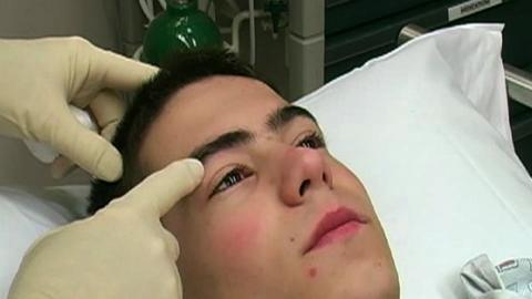
Palpate the supraorbital rim and identify the supraorbital notch (the injection site).
Cleanse the skin site with antiseptic solution, keeping it out of the eye.
Place a skin wheal (shallow intradermal injection) of anesthetic, if one is being used, at the supraorbital notch.
Insert the needle farther and gently probe medially and slightly cephalad to elicit paresthesias. Do not insert the needle into the supraorbital foramen.
When paresthesia occurs, withdraw the needle 1 to 2 mm.
Aspirate to exclude intravascular placement and then slowly (ie, over 30 to 60 seconds) inject about 3 mL of anesthetic. While injecting, apply pressure (using your finger or some gauze) under the supraorbital rim to prevent swelling of the upper eyelid.
If no paresthesia occurs during needle insertion, inject the anesthetic over the supraorbital notch (identified by palpation).
Massage the area for about 10 seconds to hasten the onset of anesthesia.
If these injections are unsuccessful, place a line of anesthetic subcutaneously along the orbital rim to block the branches of the ophthalmic nerve.
Allow about 5 to 10 minutes for the anesthetic to take effect.

Aftercare for Ophthalmic Nerve Block
Ensure hemostasis at the injection site.
Instruct patient regarding anticipated time to anesthesia resolution.
Warnings and Common Errors for Ophthalmic Nerve Block
To minimize the risk of needle breakage, do not bend the needle at its hub, insert it to its full depth (ie, to the hub), or attempt to change direction of the needle while it is inserted.
To help prevent nerve injury or intraneural injection, instruct patients to report paresthesias or pain during the nerve block procedure.
To help prevent intravascular injections, aspirate before injecting.
Tricks and Tips for Ophthalmic Nerve Block
Minimize the pain of injection by injecting slowly (eg, 30 to 60 seconds), warming the anesthetic solution to body temperature, and buffering the anesthetic with sodium bicarbonate.by injecting slowly (eg, 30 to 60 seconds), warming the anesthetic solution to body temperature, and buffering the anesthetic with sodium bicarbonate.


