

Testicular cancer begins as a scrotal mass, which is usually not painful. Diagnosis is by ultrasonography. Treatment is with orchiectomy and sometimes lymph node dissection, radiation therapy, chemotherapy, or a combination, depending on histology and stage.
In the United States, about 9760 new cases of testicular cancer and about 500 deaths (2024 estimates) occur each year (1). Testicular cancer is the most common solid cancer in males aged 15 to 35. Incidence is 2.5 to 20 times higher in patients with cryptorchidism. This excess risk is decreased or eliminated if orchiopexy is done before 10 years of age. Cancer can also develop in the contralateral normally descended testis. The cause of testicular cancer is unknown.
Most testicular cancers originate in primordial germ cells. Germ cell tumors are categorized as seminomas (40%) or nonseminomas (tumors containing any nonseminomous elements; 60%). Nonseminomas include teratomas, embryonal carcinomas, endodermal sinus tumors (yolk sac tumors), and choriocarcinomas. Histologic combinations are common; eg, teratocarcinoma contains teratoma plus embryonal carcinoma. Functional interstitial cell carcinomas of the testis are rare (< 5%) (2).
Even patients with apparently localized tumors may have occult nodal or visceral metastases. For example, almost 30% of patients with nonseminomas will relapse with nodal or visceral metastases if they undergo no treatment after orchiectomy (2). Risk of metastases is highest for choriocarcinoma and lowest for teratoma.
Tumors originating in the epididymis, testicular appendages, and spermatic cord are usually benign fibromas, fibroadenomas, adenomatoid tumors, and lipomas. Sarcomas, most commonly rhabdomyosarcoma, occur occasionally, primarily in children.

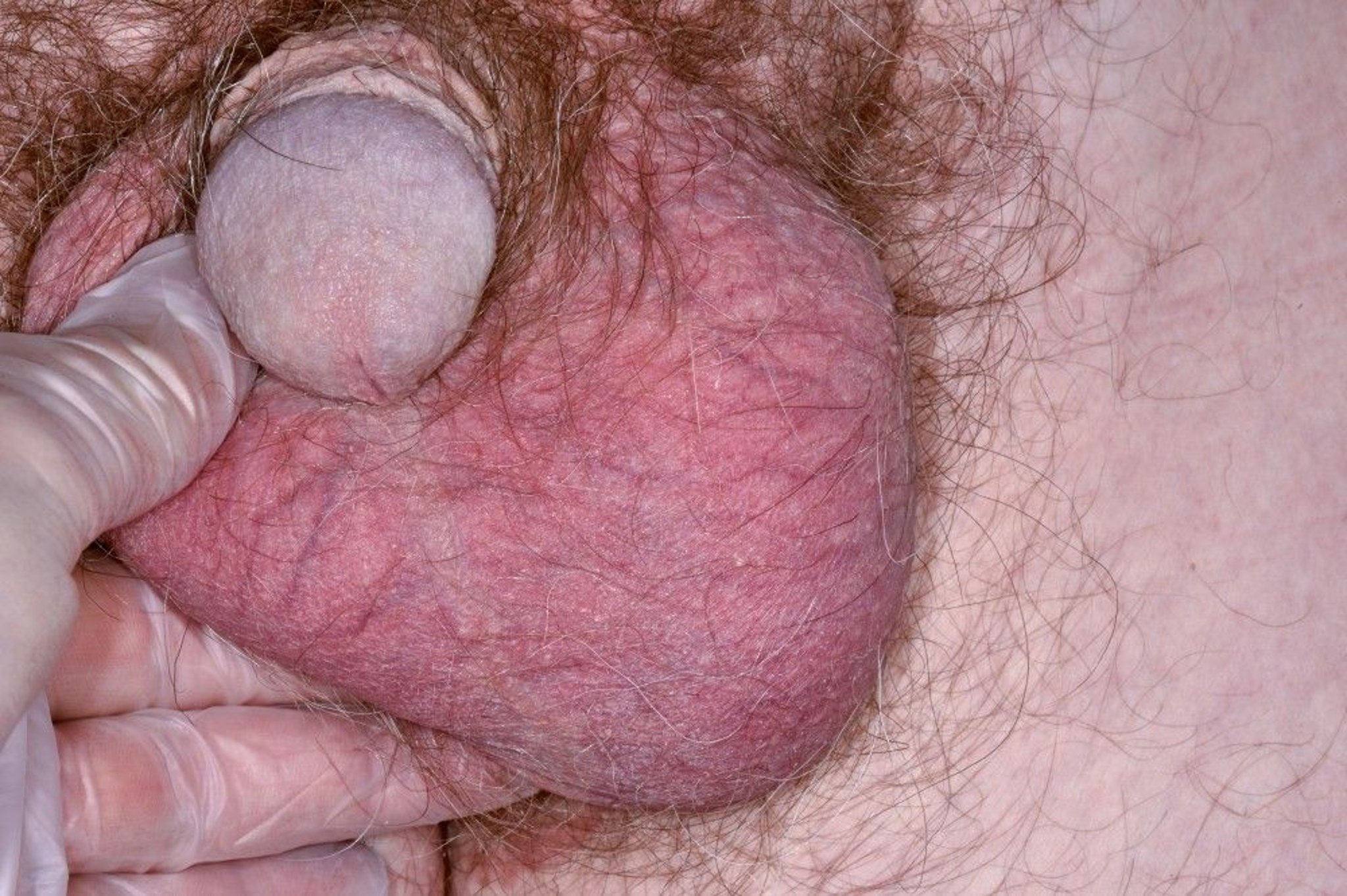
This photo shows the enlarged, painless left testis of a male patient with a seminoma.
DR P. MARAZZI/SCIENCE PHOTO LIBRARY
References
1. American Cancer Society: Key statistics for testicular cancer. Accessed February 3, 2025.
2. Goldberg H, Klaassen Z, Chandrasekar T, Fleshner N, Hamilton RJ, Jewett MAS. Germ Cell Testicular Tumors-Contemporary Diagnosis, Staging and Management of Localized and Advanced disease. Urology. 2019;125:8-19. doi:10.1016/j.urology.2018.12.025
Symptoms and Signs of Testicular Cancer
Most patients present with a scrotal mass, which is painless or sometimes associated with dull, aching pain. In a few patients, hemorrhage into the tumor may cause acute local pain and tenderness. Many patients discover the mass themselves after minor scrotal trauma. Rarely, patients with widely metastatic disease present with symptoms related to their metastases (eg, abdominal pain, low back pain, confusion or headaches, shortness of breath, chest pain).
Diagnosis of Testicular Cancer
Ultrasonography for scrotal masses
Exploration if testicular mass is present
Staging by abdominal, pelvic, and chest CT as well as tissue examination
Serum tumor markers such as AFP (alpha-fetoprotein) and beta-HCG (human chorionic gonadotropin)Serum tumor markers such as AFP (alpha-fetoprotein) and beta-HCG (human chorionic gonadotropin)
Many patients discover the mass themselves during self-examination. Monthly self-examination should be encouraged among young men.
The origin and nature of scrotal masses must be determined accurately because most testicular masses are malignant, but most extratesticular masses are not; distinguishing between the 2 during physical examination may be difficult. Scrotal ultrasonography can confirm testicular origin. If a testicular mass is confirmed, serum tumor markers (AFP, beta-HCG, and lactate dehydrogenase [LDH]) should be measured and a chest x-ray taken. Serum markers may help differentiate benign from cancerous masses but results are not definitive. Then, radical orchiectomy via an inguinal approach is indicated; the spermatic cord is exposed and clamped before the abnormal testis is manipulated.
If cancer is confirmed, abdominal, pelvic, and chest CT is needed for clinical staging using the standard TNM (tumor, node, metastasis) system (see tables and ). Tissue obtained during treatment (usually radical inguinal orchiectomy) helps provide important histopathologic information, particularly about the proportion of histologic types and presence of intratumoral vascular or lymphatic invasion. Such information can predict the risk of occult lymph node and visceral metastases. Patients with nonseminomas have about a 30% risk of recurrence despite having what appears to be localized disease. Seminomas recur in about 15% of such patients (1).
AJCC/TNM* Staging of Testicular Cancer
Stage | Tumor† | Regional Lymph Node Metastasis | Distant Metastasis | Serum Tumor Markers |
|---|---|---|---|---|
0 | pTis | N0 | M0 | S0 |
I | pT1–pT4 | N0 | M0 | SX |
IA | pT1 | N0 | M0 | S0 |
IB | pT2–pT4 | N0 | M0 | S0 |
IS | Any pT/TX | N0 | M0 | S1–S3 |
II | Any pT/TX | N1–N3 | M0 | SX |
IIA | Any pT/TX | N1 | M0 | S0, S1 |
IIB | Any pT/TX | N2 | M0 | S0, S1 |
IIC | Any pT/TX | N3 | M0 | S0, S1 |
III | Any pT/TX | Any N | M1 | SX |
IIIA | Any pT/TX | Any N | M1a | S0, S1 |
IIIB | Any pT/TX | N1–N3 | M0 | S2 |
Any pT/TX | Any N | M1a | S2 | |
IIIC | Any pT/TX | N1–N3 | M0 | S3 |
Any pT/TX | Any N | M1a | S3 | |
Any pT/TX | Any N | M1b | Any S | |
* For AJCC/TNM definitions, see table TNM and Serum Marker Definitions for Testicular Cancer. Data adapted from American Cancer Society, Testicular Cancer Stages. Accessed February 3, 2025. † pT = pathologic T-stage based on orchiectomy specimen; TX = tumor is not assessable for some reason. | ||||
TNM and Serum Marker Definitions for Testicular Cancer*
Feature | Definition |
|---|---|
Tumor | |
pTX | Not assessable |
pT0 | No evidence of primary tumor (eg, scar in testis) |
pTis | Intratubular germ cell tumors (carcinoma in situ) |
pT1 | Limited to testis and rete testis without vascular or lymphatic invasion May invade tunica albuginea but not tunica vaginalis |
pT1a | Seminoma < 3 cm |
pT1b | Seminoma ≥ 3 cm |
pT2 | Limited to testis and epididymis with vascular or lymphatic invasion, or extends through tunica albuginea and involves tunica vaginalis |
pT3 | Invades spermatic cord with or without vascular or lymphatic invasion |
pT4 | Invades scrotum with or without vascular or lymphatic invasion |
Regional lymph node metastasis | |
NX | Not assessable |
N0 | None |
N1 | ≥ 1 node but not more than 5 nodes, all ≤ 2 cm in greatest dimension |
N2 | ≥ 1 node > 2 cm but ≤ 5 cm in greatest dimension, or more than 5 nodes ≤ 5 cm in greatest dimension |
N3 | ≥ 1 node > 5 cm in greatest dimension |
Distant metastasis | |
MX | Not assessable |
M0 | None |
M1 | Present |
M1a | Nonretroperitoneal nodal or lung metastasis |
M1b | Distant metastasis other than nonregional lymph nodes or lung |
Serum markers | |
SX | Markers not available or not measured |
S0 | Levels within normal limits |
S1 | LDH < 1.5 × the upper limit of normal for the LDH assay and beta-hCG < 5000 mIU/mL and AFP < 1000 ng/mL |
S2 | LDH = 1.5–10 × upper limit of normal for the LDH assay or beta-hCG 5000–50,000 mIU/mL or AFP 1000–10,000 ng/mL |
S3 | LDH > 10 × upper limit of normal for the LDH assay or hCG > 50,000 mIU/mL or AFP >10,000 ng/mL |
* Data adapted from American Cancer Society, Testicular Cancer Stages and National Comprehensive Cancer Network ( NCCN) Clinical Practice Guidelines in Oncology. Testicular Cancer, Version 1.2023. Accessed February 3, 2025. | |
AFP = alpha fetoprotein; hCG = human chorionic gonadotropin; LDH human chorionic gonadotropin; LDH= lactate dehydrogenase; M = distant metastases; N = regional lymph nodes (assessed clinically); p = pathologic staging; S = serum tumor markers; T = main tumor. | |
Diagnosis reference
1. Kollmannsberger C, Tandstad T, Bedard PL, et al. Patterns of relapse in patients with clinical stage I testicular cancer managed with active surveillance. J Clin Oncol. 2015;33(1):51-57. doi:10.1200/JCO.2014.56.2116
Treatment of Testicular Cancer
Radical inguinal orchiectomy
Sometimes radiation, or chemotherapy for seminomas
Chemotherapy or surgery for nonseminomas
Active surveillance
Radical inguinal orchiectomy (removal of the testicle and its spermatic cord) is the cornerstone of treatment and helps provide important diagnostic information; it also helps formulate the subsequent treatment plan. A cosmetic testicular prosthesis may be placed during orchiectomy. Silicone prostheses are not widely available because of the problems with silicone breast implants. However, saline implants have been developed. For men who wish to retain reproductive capacity, sperm banking is potentially available in anticipation of radiation therapy or chemotherapy.
Lymph node dissection
For seminomas, primary retroperitoneal lymph node dissection is often done after orchiectomy in patients with nodal disease < 3 cm, but systemic therapy remains the standard of care.
For nonseminomas, many experts consider standard treatment following orchiectomy to be retroperitoneal lymph node dissection. However, for clinical stage 1 tumors in patients who have no prognostic factors that predict relapse, the recommended approach is active surveillance (frequent serum marker measurements, chest x-rays, CT). Intermediate-sized retroperitoneal nodal masses may be treated with up-front retroperitoneal lymph node dissection or chemotherapy (eg, bleomycin, etoposide, cisplatin), but the optimal sequence is undecided.For nonseminomas, many experts consider standard treatment following orchiectomy to be retroperitoneal lymph node dissection. However, for clinical stage 1 tumors in patients who have no prognostic factors that predict relapse, the recommended approach is active surveillance (frequent serum marker measurements, chest x-rays, CT). Intermediate-sized retroperitoneal nodal masses may be treated with up-front retroperitoneal lymph node dissection or chemotherapy (eg, bleomycin, etoposide, cisplatin), but the optimal sequence is undecided.
Lymph node dissection is done laparoscopically at some centers. The most common adverse effect of lymph node dissection overall is failure to ejaculate. However, a nerve-sparing dissection is often possible, particularly for early-stage tumors, which usually preserves ejaculation.
Radiation therapy
An option for seminoma after unilateral orchiectomy is radiation therapy, usually 20 to 40 grays (Gy; a higher dose is used for patients with a nodal mass) to the para-aortic regions up to the diaphragm. The ipsilateral ilioinguinal region is no longer routinely treated. Occasionally, the mediastinum and left supraclavicular regions are also irradiated, depending on clinical stage. However, in stage I disease, single-dose carboplatin has largely replaced radiation therapy due to concerns of long-term cardiovascular toxicity and higher incidence of secondary cancers and death with radiation. There is no role for radiation therapy in nonseminoma.An option for seminoma after unilateral orchiectomy is radiation therapy, usually 20 to 40 grays (Gy; a higher dose is used for patients with a nodal mass) to the para-aortic regions up to the diaphragm. The ipsilateral ilioinguinal region is no longer routinely treated. Occasionally, the mediastinum and left supraclavicular regions are also irradiated, depending on clinical stage. However, in stage I disease, single-dose carboplatin has largely replaced radiation therapy due to concerns of long-term cardiovascular toxicity and higher incidence of secondary cancers and death with radiation. There is no role for radiation therapy in nonseminoma.
Chemotherapy
Nodal masses > 5 cm, lymph node metastases above the diaphragm, visceral metastases, or persistently elevated tumor markers require initial platinum-based combination chemotherapy followed by consolidative surgery for residual masses (if tumor markers normalize with systemic therapy). Such treatment commonly controls the tumor long term. Fertility is often impaired; hence, pretreatment sperm banking should be considered. However, no risk to the fetus has been proved if pregnancy does occur.
Surveillance
Surveillance is strongly preferred for patients with stage 1 seminoma or nonseminomatous germ cell tumors after orchiectomy. It is commonly offered to patients at low risk of relapse. High-risk patients can be offered adjuvant therapy, either retroperitoneal lymph node dissections or 1 to 2 courses of chemotherapy.
Recurrences
Nonseminoma recurrences are usually treated with chemotherapy, although delayed retroperitoneal lymph node dissection may be appropriate for some patients with nodal relapse, normal tumor markers and no evidence of visceral metastases.
Prognosis for Testicular Cancer
Prognosis depends on histology and extent of the tumor. The 5-year survival rate is > 95% for patients with a seminoma or nonseminoma localized to the testis or with a nonseminoma and low-volume metastases in the retroperitoneum. The 5-year survival rate for patients with extensive retroperitoneal metastases or with pulmonary or other visceral metastases ranges from 48% (for some nonseminomas) to > 80%, depending on site, volume, and histology of the metastases, but even patients with advanced disease at presentation may be cured.
Key Points
Testicular cancer, the most common solid cancer in males aged 15 to 35 and is often curable, particularly seminoma.
Assess scrotal masses by ultrasonography and if they are testicular, do a chest x-ray and measure AFP (alpha-fetoprotein), beta-HCG (human chorionic gonadotropin) and LDH (lactate dehydrogenase).Assess scrotal masses by ultrasonography and if they are testicular, do a chest x-ray and measure AFP (alpha-fetoprotein), beta-HCG (human chorionic gonadotropin) and LDH (lactate dehydrogenase).
Primary treatment is radical inguinal orchiectomy, followed by surveillance, chemotherapy, radiation therapy, or retroperitoneal lymph node dissection.


