Pyoderma gangrenosum is a chronic, neutrophilic, progressive skin necrosis of unknown etiology often associated with systemic illness and sometimes skin injury. Diagnosis is clinical. Treatment includes wound care and, based on severity, anti-inflammatory medications or immunosuppressants.
Etiology of Pyoderma Gangrenosum
Etiology of pyoderma gangrenosum is unknown, but it can be associated with various systemic illnesses, including inflammatory bowel disease, rheumatoid arthritis, cancers, and hematologic disorders (eg, monoclonal gammopathy of undetermined significance, myelodysplastic syndrome, polycythemia vera). It is thought to be mediated by an abnormal immune response.
Most patients are ≥ 55 years of age (1).
Pyoderma gangrenosum can manifest in various subtypes.
Etiology reference
1. Xu A, Balgobind A, Strunk A, Garg A, Alloo A. Prevalence estimates for pyoderma gangrenosum in the United States: An age- and sex-adjusted population analysis. J Am Acad Dermatol. 2020;83(2):425-429. doi:10.1016/j.jaad.2019.08.001
Pathophysiology of Pyoderma Gangrenosum
Pathophysiology of pyoderma gangrenosum is poorly understood but may involve problems with neutrophil chemotaxis. Interleukin-8 is overexpressed in lesions.
Ulcerations of pyoderma gangrenosum occur after trauma or injury to the skin in about 30% of patients; this process is termed pathergy (1).
Pathophysiology reference
1. Binus AM, Qureshi AA, Li VW, Winterfield LS. Pyoderma gangrenosum: a retrospective review of patient characteristics, comorbidities and therapy in 103 patients. Br J Dermatol. 2011;165(6):1244-1250. doi:10.1111/j.1365-2133.2011.10565.x
Symptoms and Signs of Pyoderma Gangrenosum
Most often, pyoderma gangrenosum begins as an inflamed, erythematous papule, pustule, or nodule. The lesion, which may resemble a furuncle or an arthropod bite at this stage, ulcerates and expands rapidly, developing a swollen necrotic base and a raised dusky to violaceous border. An undermined border (ie, loss of underlying support tissue at the border) is common, if not pathognomonic. The ulcers can coalesce to form larger ulcers, often with cribriform or sieve-like scarring.
Systemic symptoms such as fever and malaise are common.
Symptoms and signs can vary with the subtype.
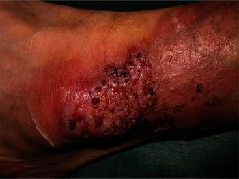
Ulcerative (classic) subtype
In this most common subtype, ulcers form as described above, most commonly on the lower extremities or trunk, particularly the buttocks and perineum.

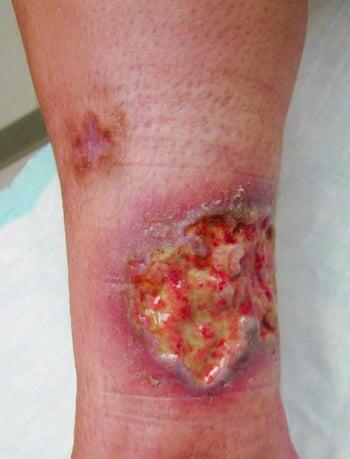


Photo courtesy of Karen McKoy, MD.
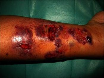
Bullous (atypical) subtype
This less common subtype often develops in patients with hematologic disorders. Lesions usually begin as bullae that erode, becoming superficial ulcers. The arms and face are most often involved.

Pustular subtype
This subtype tends to develop during exacerbations of inflammatory bowel disease. Painful pustules develop, surrounded by erythema. Arthralgias are common.
Vegetative (superficial granulomatous pyoderma) subtype
In this subtype, a single, indolent, mildly painful plaque or superficial ulcer develops, most often on the head or neck. The border is not undermined and the base is not necrotic.
Other subtypes
Pyoderma gangrenosum can also develop at other sites, such as around a stoma in patients who have inflammatory bowel disease (peristomal pyoderma gangrenosum), on the genitals (genital pyoderma gangrenosum), or in sites other than the skin, such as the bones, cornea, central nervous system, heart, intestine, liver, lungs, or muscle (extracutaneous pyoderma gangrenosum).
Diagnosis of Pyoderma Gangrenosum
Clinical evaluation
Diagnosis of pyoderma gangrenosum is clinical and is a diagnosis of exclusion after other causes of ulceration have been ruled out. Expansion of ulceration after surgical debridement strongly suggests pyoderma gangrenosum.
Biopsies of lesions are not often diagnostic but may be supportive; many biopsies from a leading edge show vasculitis with neutrophils and fibrin in superficial vessels.
Patients who have bullous (atypical) pyoderma gangrenosum should be monitored with periodic clinical assessment and complete blood count for development of a hematologic disorder.
Treatment of Pyoderma Gangrenosum
Wound care
Corticosteroids
Tumor necrosis factor (TNF)-alpha inhibitors
Sometimes other anti-inflammatory medications or immunosuppressants
Avoidance of surgical debridement
Wound healing can be promoted with moisture-retaining occlusive dressings for less exudative plaques and absorptive dressings for highly exudative plaques. Biologic and other specialized dressings may be needed in refractory cases. Wet-to-dry dressings should be avoided.
Surgical treatments are avoided because of the risk of wound extension (1).
Treatment reference
1. Alavi A, French LE, Davis MD, et al: Pyoderma gangrenosum: An update on pathophysiology, diagnosis and treatment. Am J Clin Dermatol 18(3):355–372, 2017. doi: 10.1007/s40257-017-0251-7
Key Points
Pyoderma gangrenosum is often associated with a systemic disorder and is probably immune-mediated.
There are several subtypes; the ulcerative subtype (ie, necrotic base and raised violaceous border with undermined edge on a lower extremity, buttock, or perineum) is most common.
Diagnose pyoderma gangrenosum clinically.
Optimize wound care and avoid surgical debridement.