

Osteosclerosis is a type of osteopetrosis that involves abnormal hardening of bone and increased skeletal density with little disturbance of modeling; in some types, bony encroachment on the bone marrow cavity causes cytopenias.
Osteopetroses are familial disorders caused by defective function of the osteoclasts and are characterized by increased bone density and abnormal skeletal modeling.
Osteopetrosis with Delayed Manifestations (Albers-Schönberg Disease)
Osteopetrosis with delayed manifestations is characterized by increased bone density due to defective osteoclast function.
This type of osteopetrosis is autosomal dominant, manifesting during childhood, adolescence, or young adulthood. It is less severe than osteopetrosis with precocious manifestations. The defective CLCN7 gene encodes a chloride channel that is apparently important in osteoclast function.
Osteopetrosis with delayed manifestations is relatively common, affecting approximately 1/20,000 people, and has a wide geographic and ethnic distribution (1). Affected people may be asymptomatic; general health is usually unimpaired. However, facial palsy and deafness may occur due to cranial nerve entrapment. Bony overgrowths may narrow the bone marrow cavity and cause cytopenias ranging from anemia to pancytopenia. Extramedullary hematopoiesis may then occur, resulting in hepatosplenomegaly; consequent hypersplenism can paradoxically worsen anemia.
The skeleton usually is radiologically normal at birth. However, bone sclerosis becomes increasingly apparent as children age, and diagnosis is typically based on radiographs done for unrelated reasons. Bony involvement is widespread but patchy. The calvaria is dense, and sinuses may be obliterated. Sclerosis of the vertebral end plate causes the characteristic rugby-shirt appearance (horizontal banding). Long bone fractures, scoliosis, hip osteoarthritis, and osteomyelitis of the mandible or septic osteitis or osteoarthritis elsewhere may occur (2).
Some patients require transfusion or splenectomy to treat anemia.
Osteopetrosis with delayed manifestations references
1. MedlinePlus [Internet]. Bethesda (MD): National Library of Medicine (US); [updated 2020 Jun 24]. Osteopetrosis; [updated 2010 Sep 1; cited 2025 Jun 9].
2. Sobacchi C, Villa A, Schulz A, Kornak U. CLCN7-Related Osteopetrosis. In: Adam MP, Feldman J, Mirzaa GM, Pagon RA, Wallace SE, Amemiya A, eds. GeneReviews®. Seattle (WA): University of Washington, Seattle; February 12, 2007.
Osteopetrosis with Precocious Manifestations
Osteopetrosis with precocious manifestations is a severe disorder of increased bone density due to defective osteoclast function, resulting in cranial nerve compressions, skeletal abnormalities and, ultimately, bone marrow failure.
This type of osteopetrosis is malignant, frequently lethal, and congenital, manifesting during infancy. It is autosomal recessive and often due to a mutation in the osteoclast-associated gene TCIRG1. Bony overgrowth progressively obliterates the bone marrow cavity, causing severe pancytopenia.
Osteopetrosis with precocious manifestations is rare, occurring in an estimated 1/250,000 people.
Initial symptoms of osteopetrosis with precocious manifestations include growth and weight faltering (formerly called failure to thrive), spontaneous bruising, abnormal bleeding, and anemia. Palsies of the second, third, and seventh cranial nerves and hepatosplenomegaly occur later. Bone marrow failure (anemia, overwhelming infection, or hemorrhage) usually causes death in the first year of life.
The diagnosis of osteopetrosis with precocious manifestations is suspected by the presence of bony overgrowths in the context of anemia, unusual bleeding, frequent infections, and poor growth. Typically, plain radiographs are done, along with complete blood count and coagulation tests. General increased bone density is the predominant feature on radiographs. Penetrated long bone radiographs show transverse bands in the metaphyseal regions and longitudinal striations in the shafts. As the disorder progresses, the ends of the long bones, particularly the proximal humerus and distal femur, become flask-shaped. Characteristic endobones (bone within a bone) form in the vertebrae, pelvis, and tubular bones. The skull becomes thickened, and the spine has a rugby-shirt appearance.
Hematopoietic stem cell transplantation has had excellent results. Prednisone and interferon gamma are effective in some cases.Hematopoietic stem cell transplantation has had excellent results. Prednisone and interferon gamma are effective in some cases.
Osteopetrosis with Renal Tubular Acidosis (Guibaud-Vainsel Syndrome; Marble Brain Disease)
Osteopetrosis with renal tubular acidosis is a disorder characterized by increased calcifications in the bone and brain, and kidney changes.
This type of osteopetrosis is autosomal recessive. The genetic defect involves mutations of the gene encoding carbonic anhydrase II.
Osteopetrosis with renal tubular acidosis causes weakness, stunted stature, and growth and weight faltering.
The diagnosis is based on radiographic changes of osteopetrosis (ie, bones appear dense on radiographs).Cerebral calcifications are seen. Renal tubular acidosis is present, and red blood cell carbonic anhydrase activity is decreased.
Hematopoietic stem cell transplantation may cure the osteopetrosis but has no effect on the renal tubular acidosis (1). Maintenance therapy consists of bicarbonate and electrolyte supplementation to correct renal losses.
Osteopetrosis with renal tubular acidosis reference
1. McMahon C, Will A, Hu P, Shah GN, Sly WS, Smith OP. Bone marrow transplantation corrects osteopetrosis in the carbonic anhydrase II deficiency syndrome. Blood. 2001;97(7):1947-1950. doi:10.1182/blood.v97.7.1947
Pyknodysostosis (Pycnodysostosis)
Pyknodysostosis is a rare, inherited disorder caused by mutations in the CTSK gene, leading to dense but fragile bones, short stature, characteristic facial features, dental anomalies, and a high risk of fractures.
This autosomal recessive disorder is caused by loss of function mutations in the gene encoding cathepsin K, an osteoclast-derived protease important in degradation of extracellular bone matrix.
Short stature becomes evident in early childhood; adult height is ≤ 150 cm (5 ft). Other manifestations include an enlarged skull, short and broad hands and feet, short sclerotic terminal phalanges, dystrophic nails, and retention of primary teeth. Blue sclerae (due to a deficiency in connective tissue allowing the underlying vessels to show through) are usually recognized during infancy. Affected people resemble each other closely; they have a small face, retrognathia (a receding chin), and carious, misplaced teeth. The cranium bulges, and the anterior fontanelle remains patent. Pathologic fractures are a complication of pyknodysostosis.

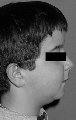
This photo shows retrognathia (a receding chin) in a boy with pyknodysostosis.
This photo shows retrognathia (a receding chin) in a boy with pyknodysostosis.
© Springer Science+Business Media

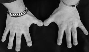
This photo shows short, broad hands and dysplasia of terminal phalanges (particularly of the thumbs) in a man with pyknodysostosis.
This photo shows short, broad hands and dysplasia of terminal phalanges (particularly of the thumbs) in a man with pykn
© Springer Science+Business Media

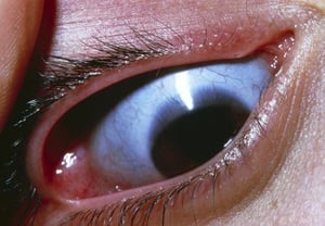
This photo shows a close-up of the eye showing a blue sclera (normally white).
This photo shows a close-up of the eye showing a blue sclera (normally white).
JAMES STEVENSON/SCIENCE PHOTO LIBRARY
This photo shows retrognathia (a receding chin) in a boy with pyknodysostosis.
This photo shows retrognathia (a receding chin) in a boy with pyknodysostosis.
© Springer Science+Business Media
This photo shows short, broad hands and dysplasia of terminal phalanges (particularly of the thumbs) in a man with pyknodysostosis.
This photo shows short, broad hands and dysplasia of terminal phalanges (particularly of the thumbs) in a man with pykn
© Springer Science+Business Media
This photo shows a close-up of the eye showing a blue sclera (normally white).
This photo shows a close-up of the eye showing a blue sclera (normally white).
JAMES STEVENSON/SCIENCE PHOTO LIBRARY
The diagnosis of pyknodysostosis is suspected by the presence of blue sclerae, short stature, and characteristic skeletal features. Typically, plain radiographs are done. Bone sclerosis appears on radiographs during childhood, but neither bone striations nor endobones (bone within bone) are seen. Facial bones and paranasal sinuses are hypoplastic, and the mandibular angle is obtuse. Clavicles may be gracile (narrower than is normal), and their lateral portions may be underdeveloped; distal phalanges are rudimentary.
Growth hormone therapy may be considered as a potential therapeutic option to improve growth. Plastic surgery has been used to correct severe deformities of the face and jaw.


