


Mastocytosis is mast cell proliferation with infiltration of skin or other tissues and organs. Mast cell activation syndrome is increased and inappropriate activation of mast cells without clonal proliferation. Symptoms result mainly from mediator release and include pruritus, flushing, and dyspepsia due to gastric hypersecretion. Diagnosis is by skin or bone marrow biopsy or both. Treatment is with antihistamines and control of any underlying disorder.
(See also Overview of Allergic and Atopic Disorders.)
Etiology in many cases of mastocytosis involves an activating mutation (D816V) in the gene coding for the stem cell factor receptor c-kit, which is present on mast cells. The result is autophosphorylation of the receptor, which causes uncontrolled mast cell proliferation.
Classification of Mastocytosis
Mastocytosis may be cutaneous or systemic.
Cutaneous mastocytosis
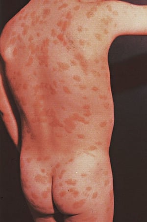
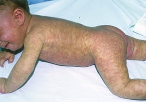
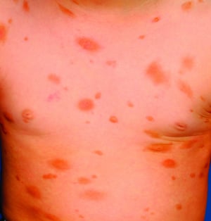
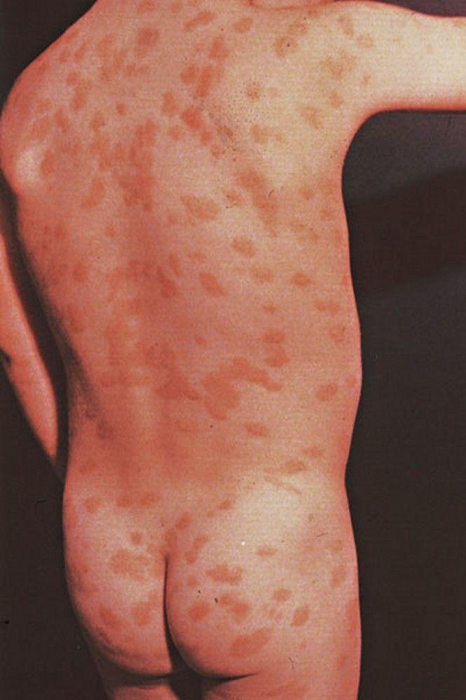
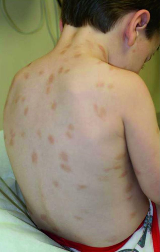
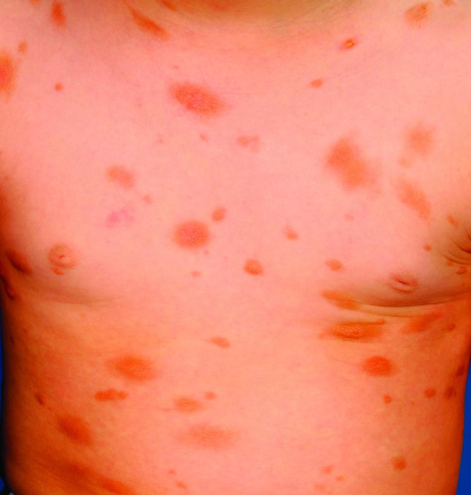
Cutaneous mastocytosis typically occurs in children. Most patients present with urticaria pigmentosa, a local or diffusely distributed salmon or brown maculopapular rash caused by multiple small mast cell collections. Nodular lesions and plaques can also develop. Less common are diffuse cutaneous mastocytosis, which is skin infiltration without discrete lesions, and mastocytoma, which is a large (1 to 5 cm) solitary collection of mast cells.
Cutaneous forms rarely progress to systemic disease in children but may do so in adults.


By permission of the publisher. From Joe E, Soter N. In Current Dermatologic Diagnosis and Treatment, edited by I Freedberg, IM Freedberg, and MR Sanchez. Philadelphia, Current Medicine, 2001.
© Springer Science+Business Media

© Springer Science+Business Media

Image courtesy of Karen McKoy, MD.
© Springer Science+Business Media

By permission of the publisher. From Joe E, Soter N. In Current Dermatologic Diagnosis and Treatment, edited by I Freedberg, IM Freedberg, and MR Sanchez. Philadelphia, Current Medicine, 2001.


© Springer Science+Business Media

© Springer Science+Business Media

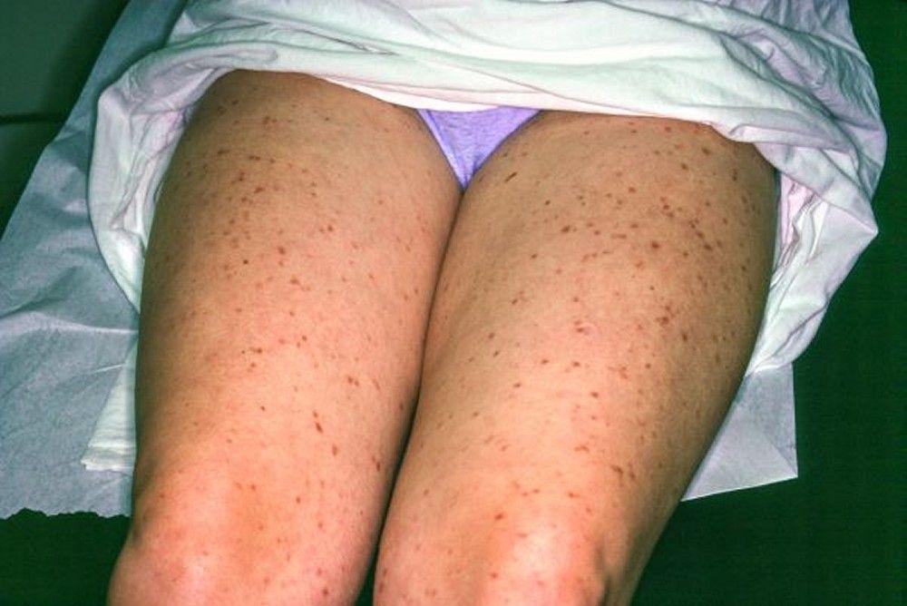
Image courtesy of Karen McKoy, MD.

© Springer Science+Business Media
Systemic mastocytosis
Systemic mastocytosis most commonly occurs in adults and is characterized by multifocal bone marrow lesions; it often involves other organs, most commonly skin, lymph nodes, liver, spleen, and/or gastrointestinal (GI) tract.
Systemic mastocytosis is classified as
Indolent mastocytosis, with no organ dysfunction and a good prognosis
Mastocytosis associated with other hematologic disorders (eg, myeloproliferative disorders, myelodysplasia, lymphoma)
Aggressive mastocytosis, characterized by impaired organ function
Mast cell leukemia, with > 20% mast cells in bone marrow, no skin lesions, multiorgan failure, and a poor prognosis
Mast cell activation syndrome
Mast cell activation syndrome is characterized by increased and inappropriate activation of mast cells with mediator release but without clonal proliferation or organ infiltration by mast cells (1). The syndrome was originally diagnosed only when mediator release was idiopathic, but it has since been expanded to include release triggered by allergen-specific IgE, certain drugs, or physical factors. Genetic causes are suspected but not proved. Most cases do not involve clonal proliferation of mast cells but are due to a lower threshold for mast cells to degranulate. Mast cell activation syndrome has frequently been associated with postural orthostatic tachycardia syndrome (POTS) and Ehlers-Danlos syndrome, although the nature of the connection is unclear.
The manifestations of mast cell activation syndrome are frequently similar to those of patients with systemic mastocytosis; they include tachycardia, syncope, urticaria, flushing, nausea, vomiting, and brain fog.
It is unclear whether mast cell activation syndrome can progress to systemic mastocytosis or another form of mast cell disease and, if so, how many patients are affected.
Classification reference
1. Weiler CR, Austen KF, Akin C, et al: AAAAI Mast Cell Disorders Committee Work Group Report: Mast cell activation syndrome (MCAS) diagnosis and management. J Allergy Clin Immunol 144 (4):883–896, 2019. doi: 10.1016/j.jaci.2019.08.023
Symptoms and Signs of Mastocytosis
Skin involvement is often pruritic in mastocytosis—whether a single mastocytoma or more diffuse disease. The following may worsen itching:
Changes in temperature
Contact with clothing or other materials
Use of some drugs, including nonsteroidal anti-inflammatory drugs (NSAIDs)
Consumption of hot beverages, spicy foods, or alcohol
Exercising
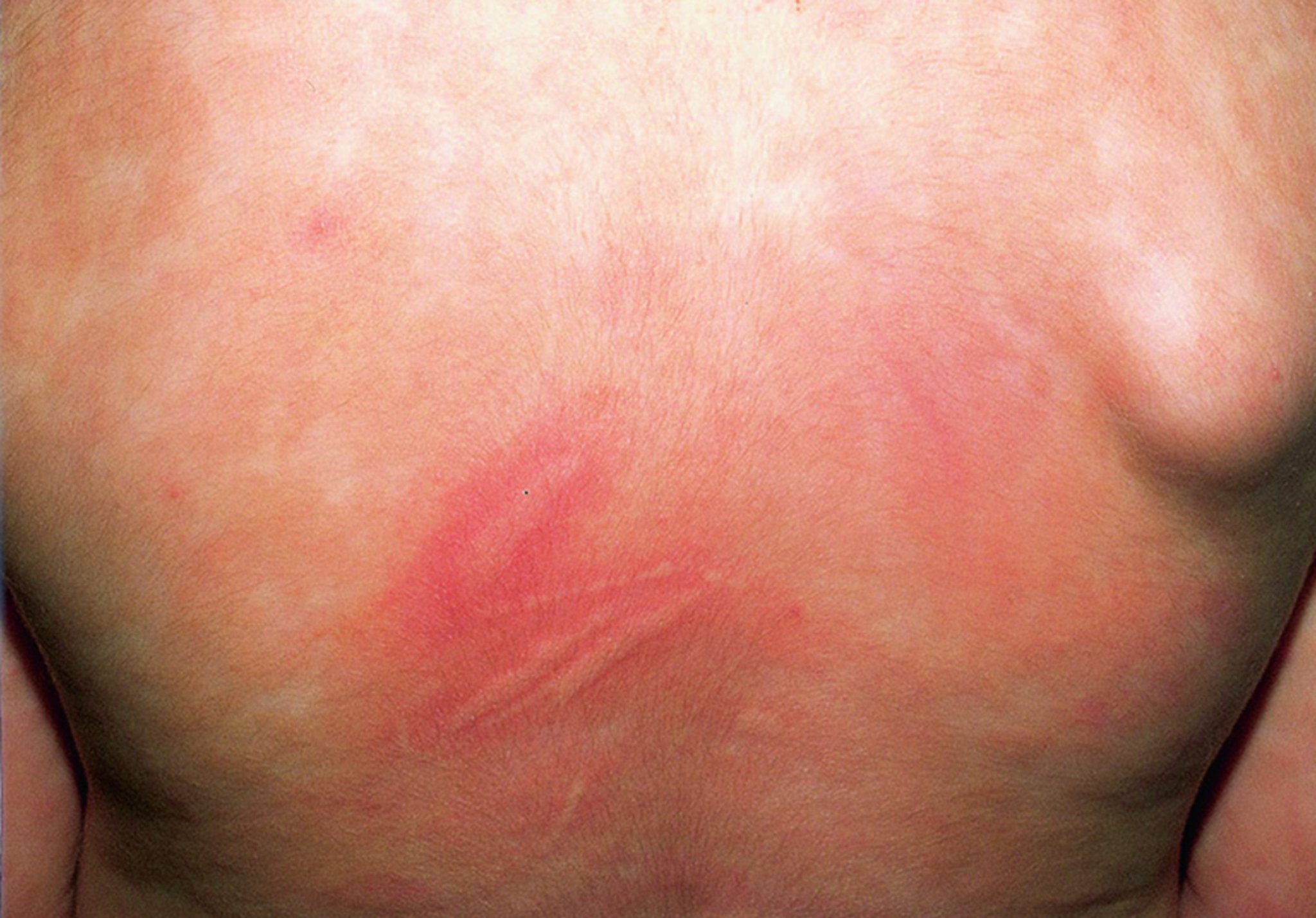
Stroking or rubbing skin lesions causes urticaria and erythema around the lesion (Darier sign); this reaction differs from dermatographism, which involves normal skin.
© Springer Science+Business Media
Systemic symptoms can occur with any form. The most common is flushing; the most dramatic are anaphylactoid and anaphylactic reactions with syncope and shock.
Other symptoms include epigastric pain due to peptic ulcer disease, nausea (because histamine stimulates gastric acid production), vomiting, chronic diarrhea, arthralgias, bone pain, and neuropsychiatric changes (eg, irritability, depression, mood lability). Hepatic and splenic infiltration may cause portal hypertension with resultant ascites.
Diagnosis of Mastocytosis
Clinical evaluation
Bone marrow biopsy
Serum tryptase levels (baseline and during symptoms if possible)
Diagnosis of mastocytosis is suggested by clinical presentation. However, similar symptoms can be caused by many other disorders such as carcinoid syndrome, vipoma, gastrinoma (Zollinger-Ellison syndrome), and chronic urticaria.
Diagnosis requires bone marrow biopsy and tryptase levels (a marker of mast cell degranulation) in most patients. Skin biopsy can be done to check for mast cells, but this test does not replace the need for a bone marrow biopsy to classify the diagnosis and staging.
Diagnosis of mastocytosis is confirmed when one major criterion and at least one minor criterion or ≥ 3 (of 4) minor criteria are met.
The major criterion is
The presence of multifocal, dense aggregates of > 15 mast cells in bone marrow (preferred) or other extracutaneous organs (except the GI tract, lymph nodes, liver, or spleen)
The 4 minor criteria are
Atypical morphology or spindle shapes in > 25% of the mast cells in bone marrow biopsy sections or aspirate
A kit mutation at codon 816 (commonly Asp816Val) in bone marrow, peripheral blood, or other tissue
Bone marrow or other extracutaneous mast cells expressing the surface markers CD2, CD25, or both
Baseline serum tryptase levels > 20 ng/mL (> 20 mcg/L); values > 11.4 ng/mL (11.4 mcg/L) are considered elevated in most diagnostic laboratories)
The baseline level of tryptase is elevated in systemic mastocytosis but is typically normal in cutaneous mastocytosis and mast cell activation syndrome.
Diagnostic criteria for mast cell activation syndrome include all of the following:
Typical clinical symptoms of mast cell mediator release
During symptoms, a substantial transient increase in total serum tryptase level or an increase in other mast cell–derived mediators, such as N-methyl histamine or prostaglandin D2, leukotriene E4, or their urinary metabolites
A clinical response to drugs that attenuate the production or activities of mast cell mediators
Patients with mast cell activation typically have a normal bone marrow biopsy if it is done.
If the diagnosis is uncertain, levels of mast cell mediators and their metabolites (eg, 24-hour N-methylhistamine, prostaglandin D2, leukotriene E4) may be measured in plasma and urine; elevated levels support the diagnosis of mast cell disease but not necessarily systemic mastocytosis.
A bone scan and GI evaluation, can also be helpful in cases where the diagnosis requires confirmation.
Diagnosis reference
1. Adapted from Horny HP, Akin C, Arber DA, et al: Mastocytosis. In WHO (World Health Organization) Classification of Tumors of Hematopoietic and Lymphoid Tissues, edited by SH Swerdlow, E Campo, and NL Harris, et al, Lyon, IARC (International Agency for Research on Cancer) Press, 2017, p. 62.
Treatment of Mastocytosis
For cutaneous mastocytosis, H1 blockers and possibly psoralen plus ultraviolet light or topical corticosteroids
Cutaneous mastocytosis
H1 blockers are effective for symptoms. Children with cutaneous forms require no additional treatment because most cases resolve spontaneously.
Adults with cutaneous forms may be treated with psoralen plus ultraviolet light or with topical corticosteroids once or twice a day.
Mastocytoma usually involutes spontaneously and requires no treatment.
Systemic mastocytosis
Management of anaphylactic reactions
1) can be used to help control end-organ damage, cytopenias, and mast cell accumulation in bone marrow (2). Dosages are
c-kit mutation.
Mast cell activation syndrome
Known triggers should be avoided.
Treatment references
1. Radia D, Deininger M, Gotlib J, et alKIT D816V, induces complete and durable responses in patients (pts) with advanced systemic mastocytosis (AdvSM). EHA Library. 2019;267413:S830.
2. Gotlib J, Kluin-Nelemans HC, George TI, et alN Engl J Med 374 (26):2530–2541, 2016. doi: 10.1056/NEJMoa1513098
Key Points
Patients with cutaneous mastocytosis, usually children, typically present with a diffuse salmon or brown, often pruritic maculopapular rash.
Systemic mastocytosis causes multifocal bone marrow lesions, usually in adults, but often affects other organs.
All types can cause systemic symptoms (most commonly, flushing but sometimes anaphylactoid reactions).
For cutaneous mastocytosis, use H1 blockers to relieve symptoms, and in adults, consider treatment with psoralen plus ultraviolet light or topical corticosteroids.
For mast cell activation syndrome, target mediators released during activation with antihistamines, leukotriene inhibitors, and mast cell stabilizers.


